Category: Diagnostic Imaging
A Look at Palm Beach Equine Clinic’s Advanced Imaging Technology
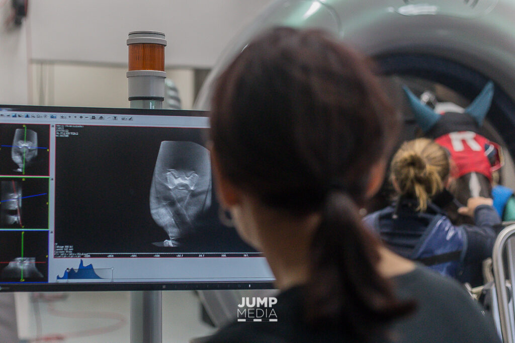
Photo by Jump Media
Palm Beach Equine Clinic takes pride in being a world-class facility for the diagnosis, treatment, and recovery of some of the industry’s most valued sport horses, as well as backyard companions. A vital area of the Equine Hospital that helps veterinarians to diagnose subtle or acute lameness is the Advanced Imaging Department.
Producing thousands of scans a year across all modalities, the Palm Beach Equine Clinic Imaging Department offers an on-staff, board-certified radiologist, and equipment that elevates the effectiveness of lameness diagnostics. Lameness is no longer a guessing game as PBEC veterinarians have an arsenal of imaging technologies to capture inside the horse.
Tour the PBEC Imaging Department:
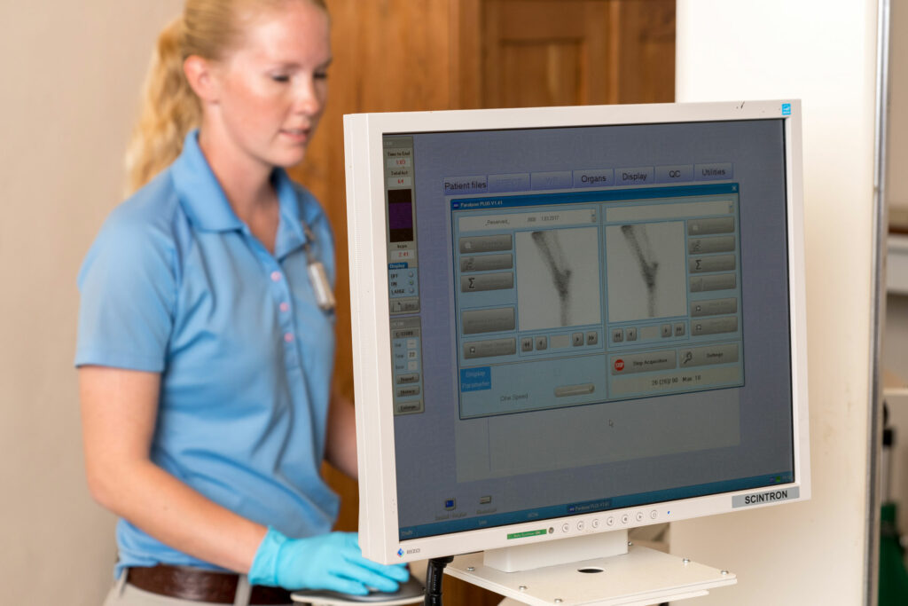
Nuclear Scintigraphy (Bone Scan)
Nuclear scintigraphy begins with the injection of a radioactive isotope called Technetium 99 that is bound to a phosphate analogue. The isotope – phosphate molecule attaches to the mineral matrix of the bone in areas where bone is active. A gamma camera is then used to capture images of the skeletal anatomy. Points that “light up” on the image indicate increased metabolic activity as a possible site of injury.

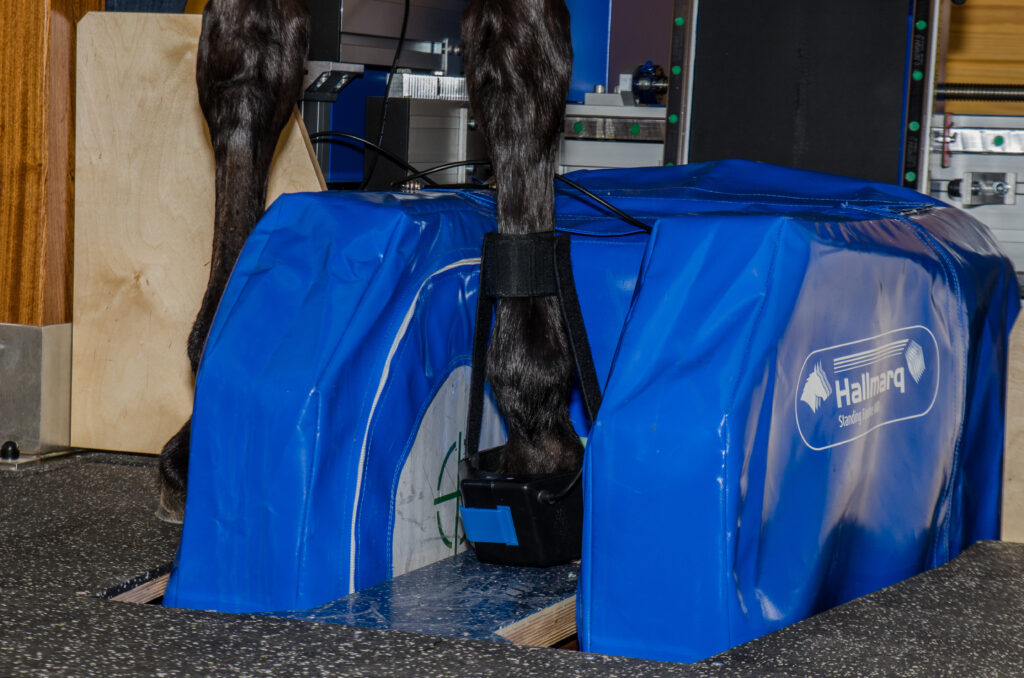
Standing Magnetic Resonance Imaging (MRI)
The standing MRI produces highly detailed, cross-sectional images of bone and soft tissue in multiple different planes to fully image a desired region. The standing MRI requires sedation (not anesthesia) and is best used to further define a specific area that has already been pinpointed as the origin of lameness.
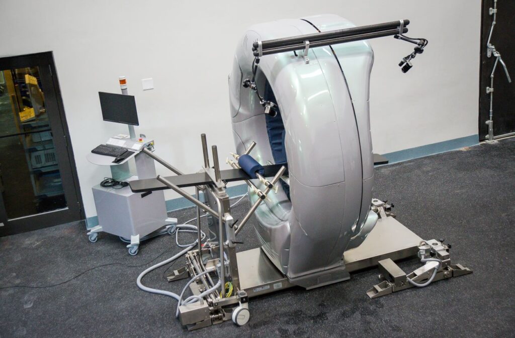
Computed Tomography (CT)
Similar to its use in humans, CT allows veterinarians the unique opportunity to conveniently explore areas of a horse’s body that were previously inaccessible. The machine produces 360 degree images of a horse’s neck, spine, and head and can be conducted while a horse is standing and under only light sedation.
Endoscopy
An endoscope is an instrument that allows veterinarians to look inside the body by being inserted through a natural opening or incision. A tiny camera on the instrument allows an in-depth view of internal structures such as the upper and lower respiratory track, gastrointestinal and urinary tracts, as well as the cervix and uterus of mares.

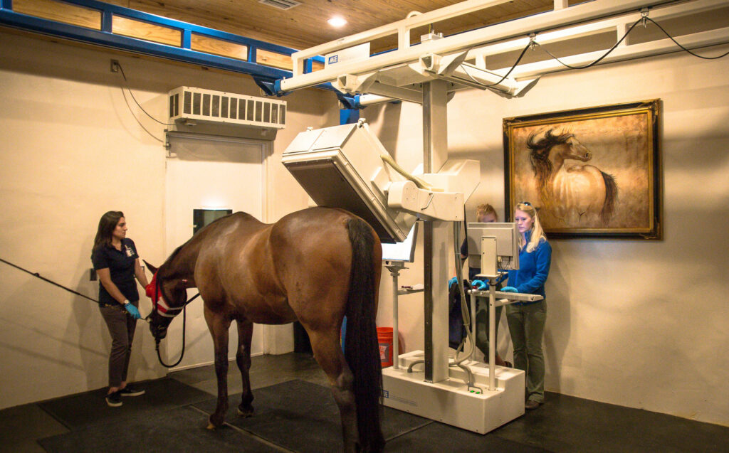
Digital Radiography
Used routinely, radiographs are traditional x-rays that are made available within seconds for digital viewing and evaluation.
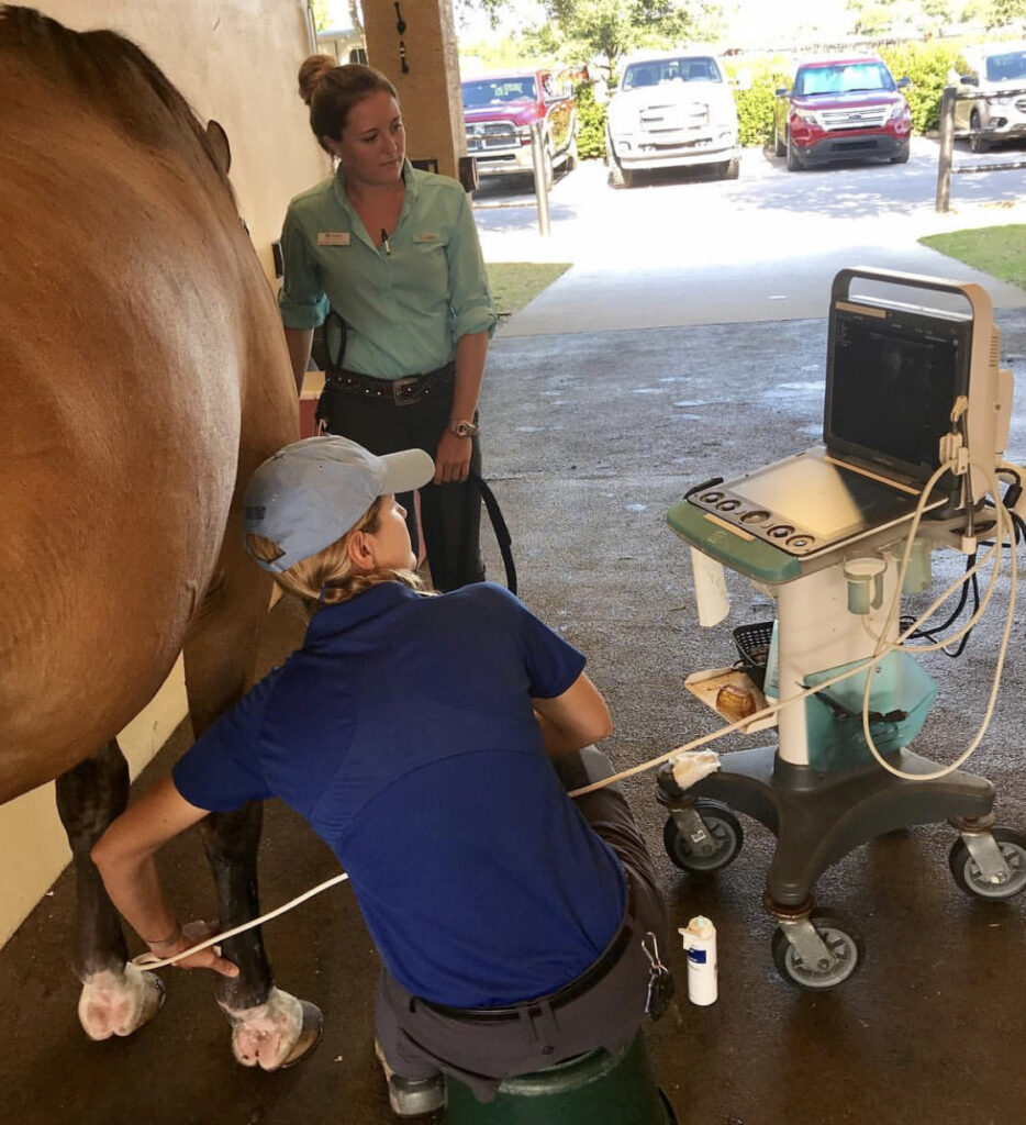
Ultrasonography
An ultrasound machine generates high-frequency sound waves, which echo an image back to the machine where bone appears white, fluid appears black, and all other structures are on a grayscale. An ultrasound is non-invasive, usually does not require sedation, does not use radiation or require injecting radioactive isotopes, and provides real-time images.
“These tools can give a definitive diagnosis, and that saves time and money in the long run,” said board-certified radiologist Dr. Sarah Puchalski. “For example, if a horse goes lame and it gets seen and treated empirically, which is a diagnosis based on likely problems through common diagnostic procedures, it either stays sound or it becomes lame again or even non-functional in three to six months. This method sets back the commencement of the appropriate therapy.
“What these modalities do is allow the horse to be treated early and correctly,” continued Dr. Puchalski. “Otherwise, you may not be treating the correct issue, and the horse could end up lame again very soon.”
Brittany Riddle, manager of PBEC’s Nuclear Scintigraphy Department, is fascinated by the structure of the horse’s body and helps to produce hundreds of bone scans per year.
“I’ve always had a strong interest in the anatomy of horses,” said Riddle, who spends her workday keeping patients comfortable and calm while operating a gamma ray camera housed in its own suite at Palm Beach Equine Clinic. “The horses are under light sedation for standing scans and these usually take from one to four hours depending on the type of scan. Usually during the winter competition season, we have anywhere from two to three horses being scanned daily.
“The variety of patients we treat is always interesting,” continued Riddle. “We see patients from all aspects of the industry. From racehorses to polo ponies, western performance, dressage, top show jumpers and hunters.”
According to Palm Beach Equine Clinic President Dr. Scott Swerdlin, having the latest in diagnostic capabilities drives the success of the Clinic.
“Many years ago, we committed to establishing Palm Beach Equine Clinic as an all-inclusive hospital and making it the most advanced referral center in the country,” said Dr. Swerdlin. “By investing in the very best personnel and equipment, we are able to provide the very best care we can for our patients.”
Typically, when a horse’s gait feels off or may be lacking usual impulsion, the rider often assumes it to be an issue of lameness associated with the forelimbs or hindlimbs. However, that may not always be the case. Utilizing advanced diagnostic imaging techniques, Palm Beach Equine Clinic is able to accurately pinpoint the specific area that is affecting overall performance. In many cases, the cervical vertebrae are often identified as the cause of lameness, asymmetry, and poor performance.
Vertebral Anatomy

The neck is composed of seven articulating cervical vertebrae running from the head to the thorax, named C1 through C7. The neck allows movement of the head while protecting the spinal cord and providing an avenue for nerves to travel. Impingement on the spinal cord and nerves connected to the cervical vertebrae can exhibit neurologically as ataxia, neck pain, or lameness.
Signs of Lameness Related to the Neck
In a lameness exam, a veterinarian will perform flexion tests and palpate areas of the body looking for decreases in the horse’s range of motion or pain upon flexion. The rider may pick up on subtle lameness issues associated with the neck by feeling a change in the horse’s suppleness or resistance to yielding in a certain direction. Lameness may even present itself as a difference in the horse’s balance, such as being heavier on the forehand, or performance issues such as late lead changes. The tried-and-true “carrot test” can also show if a horse is resistant to flexing their neck.
Identifying Lameness through Diagnostic Imaging
Historically, neck issues related to performance are generally diagnosed through a process of ruling out other areas of the body. Diagnostic imaging can now be the most powerful and effective tool for identifying the cause of lameness related to cervical injury and hereditary malformation.
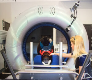
Computed Tomography (CT) scans have revolutionized the ability to assess the entire neck and can be performed while the horse is standing and under light sedation. Computed Tomography images can be rendered into three-dimensional models and sliced in any orientation, allowing the veterinarian to evaluate the vertebrae in great detail that is incomparable to standard radiographs (x-rays). These comprehensive CT scans offer veterinarians a thorough profile so they can accurately diagnose and initiate an effective response.
A standing CT scanner is the latest addition to Palm Beach Equine Clinic’s arsenal of diagnostic imaging modalities. Currently, Palm Beach Equine Clinic is the only equine hospital in South Florida offering this capability. Compared to other modalities such as MRI or Nuclear Scintigraphy, Computed Tomography offers a valuable return for its rapid acquisition of images. If you suspect there is an issue in your horse’s neck please, contact Becky at Palm Beach Equine Clinic at 561-793-1599 to schedule an appointment.
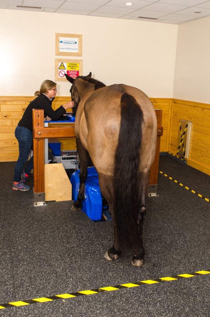
Palm Beach Equine Clinic has the most advanced state-of-the-art diagnostic imaging equipment available. More specifically, Equine Standing Magnetic Resonance Imaging (MRI) allows Palm Beach Equine Clinic to quickly and accurately diagnose injuries for their clients.

Every horse owner dreads seeing signs of lameness or discomfort in any horse, whether it is a backyard companion or a top-caliber sport horse. For performance horses, however, one of the first questions many owners ask upon contacting a veterinarian about a problem is, “Can the horse safely and comfortably return to work?” Using Palm Beach Equine Clinic’s cutting-edge equine standing MRI technology, the clinic veterinarians are best equipped to answer that question.
The equine standing MRI produces highly detailed images in several different planes to capture a complete image of the desired area. An MRI is best used to further define a specific area of bony or soft tissue that has been pinpointed as the origin of lameness. The process can be completed while the horse is in a standing position and requires only light sedation.

Lameness or performance problems are most frequently approached through routine x-rays and ultrasounds, which can come back normal. Thus, it is difficult to diagnose subtle problems because the most common tools are not sensitive enough to pick them up. At Palm Beach Equine Clinic, the Equine Standing MRI gives veterinarians an advantage when troubleshooting a lameness issue and helps them to determine a correct diagnosis in a timely manner.
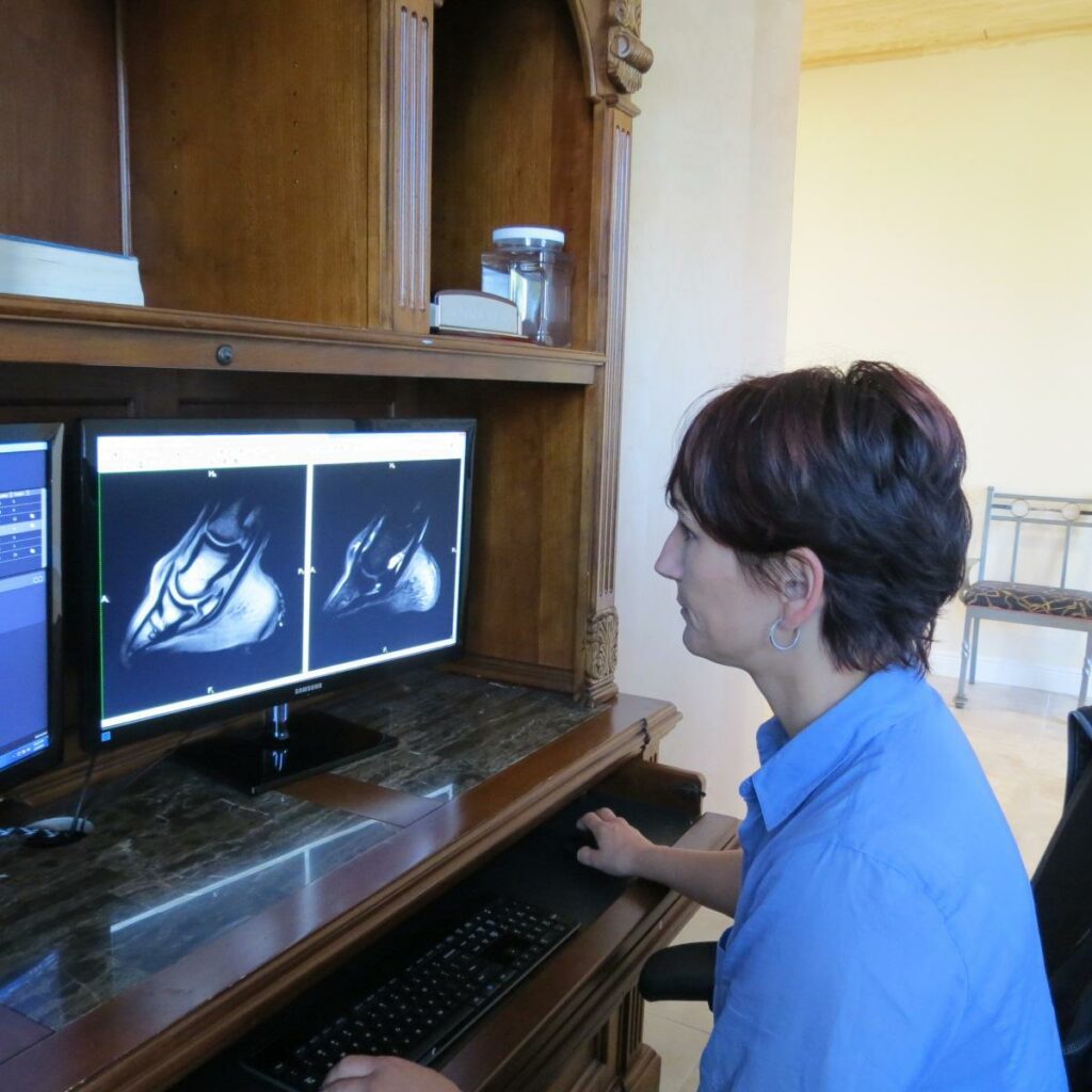
Hundreds of MRIs are read each year at Palm Beach Equine Clinic. In addition to being a state-of-the-art diagnostic tool, the equine standing MRI technology also affords economic benefits to owners by having their horse’s problem diagnosed and treated safely, effectively, and quickly.
Palm Beach Equine Clinic in Wellington, FL, has state-of-the-art surgical and advanced diagnostic imaging equipment available. With board-certified Radiologist Dr. Sarah Puchalski, Palm Beach Equine Clinic uses the Equine Standing Magnetic Resonance Imaging (MRI) and a Nuclear Scintigraphy (bone scan) camera to quickly and accurately diagnose injuries for their clients.
Every horse owner dreads seeing signs of lameness or discomfort in any horse, whether it is a backyard companion or a top-caliber sport horse. For performance horses, however, one of the first questions many owners ask upon contacting a veterinarian about a problem is, ‘Can the horse safely and comfortably return to work?’ Using Palm Beach Equine Clinic’s cutting-edge technology, Dr. Puchalski can quickly and accurately answer that question.
The Equine Standing MRI produces highly detailed images in several different planes to capture a complete image of a desired area. An MRI is best used to further define a specific area of both bony or soft tissue that has been pinpointed as the origin of lameness. The process can be completed while the horse is in a standing position and requires only light sedation.
Similarly, the process of Nuclear Scintigraphy (bone scan) begins with the injection of a radioactive isotope, specifically named Technetium 99. The isotope attaches to the phosphorous proteins localized within the bone and is absorbed over a few hours’ time. A specialized nuclear isotope gamma ray camera is used to capture images of the skeletal anatomy with a 360-degree view. Points of interest “light up” on the image to indicate increased metabolic activity and the site of injury.
Lameness or performance problems are most frequently approached through routine x-rays and ultrasounds, which can appear normal. Thus, it is difficult to diagnose subtle problems because the most common tools are not sensitive enough to diagnose in every case. At PBEC, the Equine Standing MRI and Nuclear Scintigraphy equip veterinarians with an advantage when troubleshooting a lameness issue and helps them to determine a correct, quick diagnosis.
Coupled with advanced technology, Palm Beach Equine Clinic is also one of very few equine practices in the U.S. with a Board Certified Radiologist on staff, and thanks to Dr. Puchalski, hundreds of MRI and bone scans are read each year at Palm Beach Eqine Clinic. In addition to state-of-the art diagnostic tools, the technology also affords economic benefits to owners.
“MRIs can give a definitive diagnosis, and that saves time and money in the long run,” said Dr. Puchalski. “For example, if a horse goes lame and is examined and treated empirically, which is a diagnosis based on likely problems through common diagnostic procedures, it either stays sound or it becomes lame again or even non-functional in three to six months. This method sets back the commencement of the appropriate therapy.
“What the MRI does is allow the horse to be treated early and correctly,” continued Dr. Puchalski. “Otherwise, you may not be treating the correct issue, and the horse could end up lame again very soon.”
Nuclear Scintigraphy does not produce a scan that is as specific, but it gives Dr. Puchalski the opportunity to procure a concrete quick diagnosis, as well as evaluate the whole horse for secondary problems.
“Oftentimes the primary problem in one place is making a horse sore in other places,” she said. “Owners like to know the root problem, but to also quickly diagnose secondary problems so the entire horse can be treated at once.”
As the official veterinary hospital of the Winter Equestrian Festival (WEF) and the Adequan® Global Dressage Festival (AGDF), Palm Beach Equine Clinic sees a high concentration of sport horses in need of care. In turn, owners of those horses are eager to see their horses quickly and happily return to competition.
“The biggest benefit to Palm Beach Equine Clinic and the Wellington community as a result of these MRI and Nuclear Scintigraphy scans is accessibility,” concluded Puchalski. “Anyone can call from the horse show to the clinic, get a scan scheduled quickly- in and out, get results fast, and then their training program can be changed immediately.”
About Dr. Sarah Puchalski
Dr. Sarah Puchalski is from Davis, CA, where she was an associate professor at the University of California in their Department of Surgical and Radiological Sciences. In 1995, she received her BS in biology from Simon Fraser University in British Columbia, and in 1999 earned her Doctor of Veterinary Medicine from the University of Saskatchewan in Saskatoon, where she received the ACVS Outstanding Large Animal Surgery Student award that same year. Dr. Puchalski interned in Field Service and Sports Medicine at New Bolton Center at the University of Pennsylvania in 2001, and completed her residency in radiology at UC Davis in 2005.
Dr. Puchalski has devoted her career to teaching and improving equine health through the development and refinement of diagnostic techniques. In 2011, she contributed to two books on the topic of equine lameness. Her recent contributions include chapters in “Diagnosis and Management of Lameness in the Horse,” edited by Ross and Dyson, as well as in “Veterinary Computed Tomography and the Clinical Veterinary Advisor: The Horse, Equine Colic and Veterinary Clinics of North America.” She also has contributed close to 50 scientific articles concerning the diagnosis of equine lameness to many periodic journals, including Veterinary Radiology & Ultrasound: the official journal of the American College of Veterinary Radiology and the International Veterinary Radiology Association; Veterinary Pathology; Equine Veterinary Journal; the American Journal of Veterinary Research; Equine Veterinary Education; Journal of the American Veterinary Medical Association; and Journal of Veterinary Internal Medicine.
Palm Beach Equine Clinic provides experience, knowledge, availability, and the very best care for its clients. Make Palm Beach Equine Clinic a part of your team!
Palm Beach Equine Clinic prides itself as being a consistent leader in sport horse medicine and continues to expand the diagnostic imaging technologies to provide the best services for clients. In addition to state-of-the-art imaging technology available on-site, Palm Beach Equine Clinic is fortunate to work directly with world-renowned Board Certified Radiologist Dr. Sarah Puchalski.
Dr. Sarah Puchalski is a Diplomate of the American College of Veterinary Radiology whose specialty includes the interpretation of diagnostic imaging including Radiographs, MRIs, Nuclear Scintigraphy, and CT scans. Dr. Puchalski’s job requires a high level of specialization to properly review imaging to produce comprehensive written reports for referring veterinarians. In addition to her full-time position with PBEC, Dr. Puchalski reads imaging cases for clinics all over the world. Many veterinarians and owners request a consultation as a second opinion on Pre-Purchase examinations radiographs and ultrasound evaluations.
Pioneering Equine Diagnostic Imaging Modalities
Palm Beach Equine Clinic has always been a pioneer in advances of technology within the equine veterinary industry. Almost 30 years ago, Palm Beach Equine Clinic bought the first ultrasound for equine practice in South Florida. Twenty-five years ago, Palm Beach Equine Clinic installed the first Nuclear Scintigraphy gamma ray camera to perform bone scans. Twenty years ago, Palm Beach Equine Clinic assisted in developing Computed Radiography (CR) for horses. Currently, Palm Beach Equine Clinic has the most advanced surgical and diagnostic imaging equipment available, including a standing MRI unit, MiE gamma ray camera, Digital Radiography, Video Endoscopy, and a bevy of additional diagnostic equipment.
“Palm Beach Equine Clinic has a great case population and great equipment, which is a huge bonus for someone doing what I do,” Dr. Puchalski stated. “The equipment is exceptional, the technical staff is excellent, and the case population of the region is obviously amazing.”
Nuclear Scintigraphy Imaging
Palm Beach Equine Clinic proudly offers an updated Nuclear Scintigraphy lab (bone scan) that houses the MiE Nuclear Scintigraphy gamma ray camera. Nuclear Scintigraphy is typically used to diagnosis injuries or bone remodeling within the skeletal anatomy of the horse. This specialized camera is equipped with sharper contours for precise imaging that results in accurate lameness diagnoses. Advanced software provides the ability to acquire high quality images despite small movements from the patient. This feature reduces the time required to complete a study, which provides quicker results.
Bone scans are also very useful in defining multi-limb lameness origins for the hard to diagnose, long-duration lameness cases. Typically, Nuclear Scintigraphy scans isolate points of injury to be identified further with other diagnostic techniques, such as digital radiology and magnetic resonance imaging (MRI).
The clinic offers an MRI lab containing the innovative Equine Standing MRI manufactured by Hallmarq, which scans the equine distal limb in a standing position requiring only light sedation. MRI is very useful to further define a suspected lameness origin by acquiring more defined images of boney and soft tissue structures.