Neuromuscular Biopsies: Cracking the Code on Muscle Disorders in Horses
Identifying the root of performance issues in equine athletes can be like connecting a puzzle of often-ambiguous signs, or a process of elimination through diagnostics. Veterinarians can exhaust all the tools in their medical arsenal trying to figure out why a horse may be losing muscle mass and stamina or refusing to work, but the answer could be hiding in the horse’s muscle tissue. Palm Beach Equine Clinic’s Internal Medicine Specialist Peter Heidmann, DVM, DACVIM, has a keen interest in connecting these signs and identifying muscle disorders.
“Correctly diagnosing and treating muscle disorders in horses can be a very rewarding process, because once you develop an effective treatment plan, the horse will usually experience an extraordinary recovery. I’ve had cases where we sent muscle biopsy samples to a pathologist, enacted a treatment plan, and then sent another sample six months later to check our progress. The results were so impressive that the pathologist called to confirm that it was the same horse!”
Dr. Heidmann
Muscle disorders can present with a range of signs from muscle stiffness and pain to extreme cases of muscle atrophy. Most commonly, presentations occur during training and may include pain, stiffness, tremors, and a reluctance to move. Such symptoms usually lead veterinarians to conduct a full lameness evaluation.
“We usually look at mechanics first, and when there is no lameness determined and no joint, tendon, or ligament issues, we can further narrow down the search to determine if there is nerve impingement leading to muscle atrophy,” said Dr. Heidmann, who will often see a horse with such impingement work very well at an extended trot, but lose all power or appear stiff and lame as soon as the gait is collected.
To confirm a horse’s neuromuscular health and proper function, Dr. Heidmann will first run a blood panel to test for abnormal elevations in two enzymes. The first is serum creatine kinase (CK), which is released within just a few hours of muscle damage. Elevations in CK are usually consistent with training, transport, or taxing exercise. The second is serum aspartate transaminase (AST), which is an enzyme that rises more slowly after muscle injury, and can provide a veterinarian with knowledge about the body’s longer-term response.
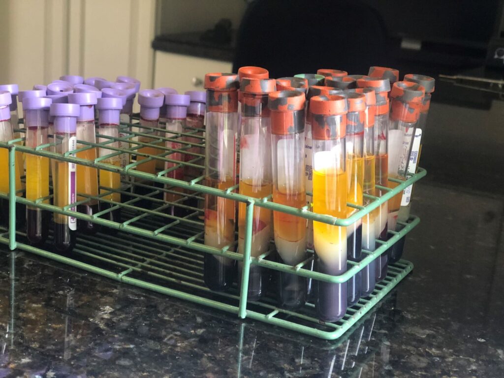
“If you think of muscle cells as little balloons, once you pop one, CK and AST are released into the blood,” said Dr. Heidmann. “When these numbers are high, we know we have way too many balloons popping all at the same time. Then we just have to figure out why.”
Neuromuscular Biopsies Leading to Diagnoses
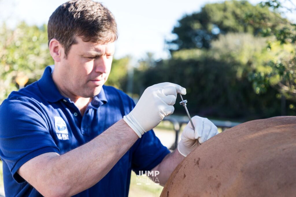
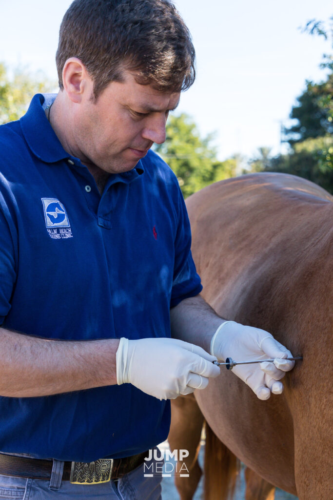
The diagnosis of a particular neuromuscular disorder can be challenging, since many of the symptoms are the same, regardless of the underlying disease. After blood testing, neuromuscular disorders can be diagnosed using a muscle biopsy. This minimally invasive, but highly revealing procedure is one that Dr. Heidmann turns to often in order to not only diagnose, but to also provide life and career-saving treatments.
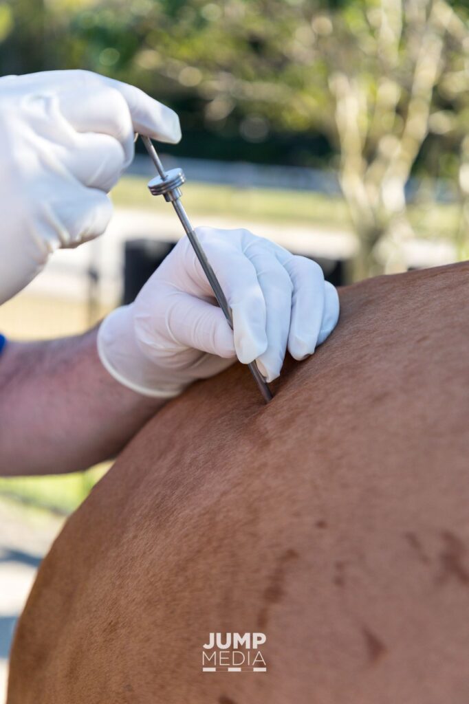

“I usually use a Bergstrom needle, which is used to collect the sample through a tiny site in the horse’s skin. The muscle tissue then falls into a slot in the needle, and is removed to be examined under a microscope,” said Dr. Heidmann, who also says the most common locations for collecting muscle biopsies, depending on the symptoms, include the triceps, top line, hamstring, gluteal muscle, and tail head of a horse. “You can physically see some of the things that aren’t functioning properly even if the enzymes aren’t high.
“The beauty of a muscle biopsy is that you are looking at where the nerves come into the muscle, so you can detect abnormal nerve supply to an area while leaving the horse with nothing more than a nick in the skin,” he continued. “Once we see the muscle tissue, we can tell if the horse requires nutritional changes, injections, or other treatments. Non-invasive nerve stimulation is what the future holds for the treatment of many muscle disorders, but it is still an evolving treatment in veterinary medicine right now.”
Once the muscle biopsy is collected, Dr. Heidmann can go to work identifying the problem while the horse immediately returns to work. Non-invasive diagnostics and treatments are the wave of the future in both human and equine medicine. For Dr. Heidmann, neuromuscular biopsies are helping him to look below the surface of a horse’s skin to evaluate, diagnose, and provide treatments without making a single cut.
Learn more about Equine Internal Medicine by clicking here.
Appointment Request
Please fill out this form and our staff will contact you to confirm an appointment.