Show Jumper Recovers from Infectious Pedal Osteitis
A 12-year-old show jumper is moving nicely just 2 months after laboring to walk.
Palm Beach Equine Clinic Staff Surgeon Dr. Weston Davis operated on a severe lameness case on July 5 that had quickly progressed into an emergency situation.
Evaluation and Diagnosis
Annabelle was referred to Palm Beach Equine Clinic for advanced imaging and evaluation of severe subsolar abscessation in the Holsteiner mare’s left front foot that was not responding to aggressive therapies.
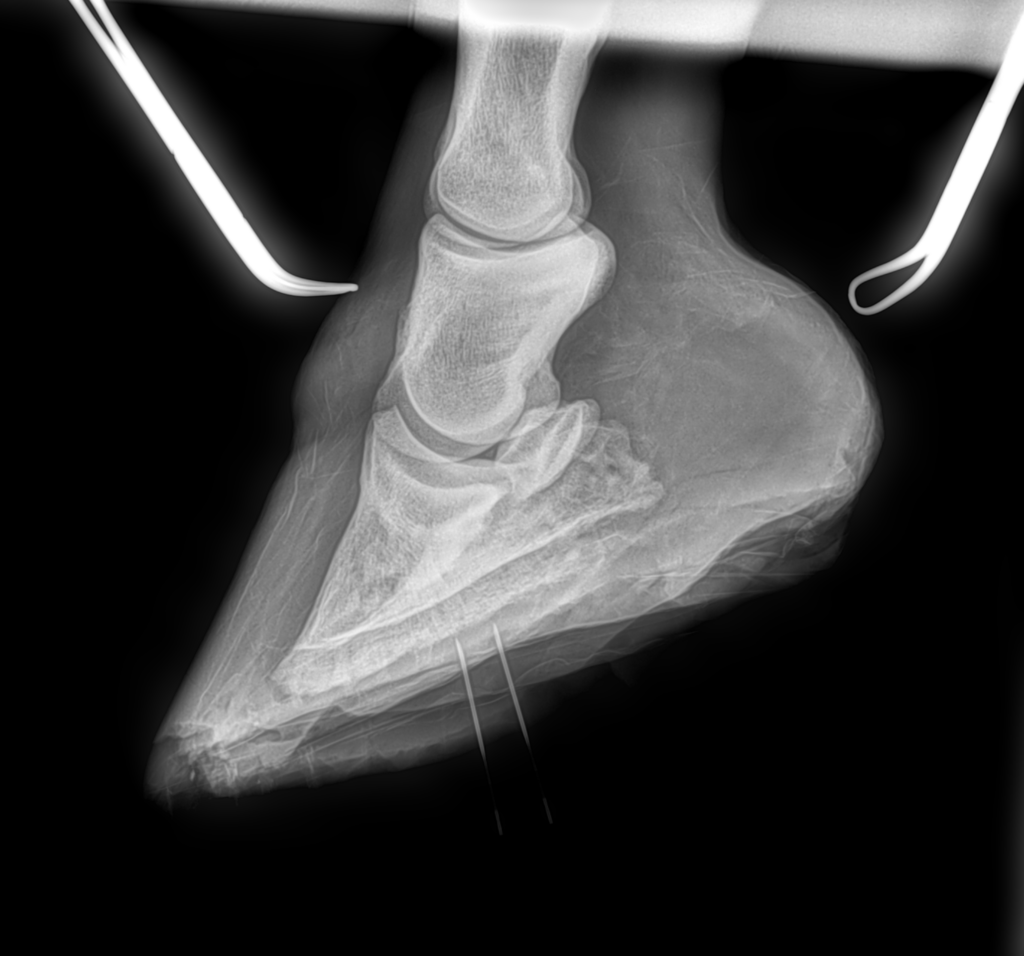
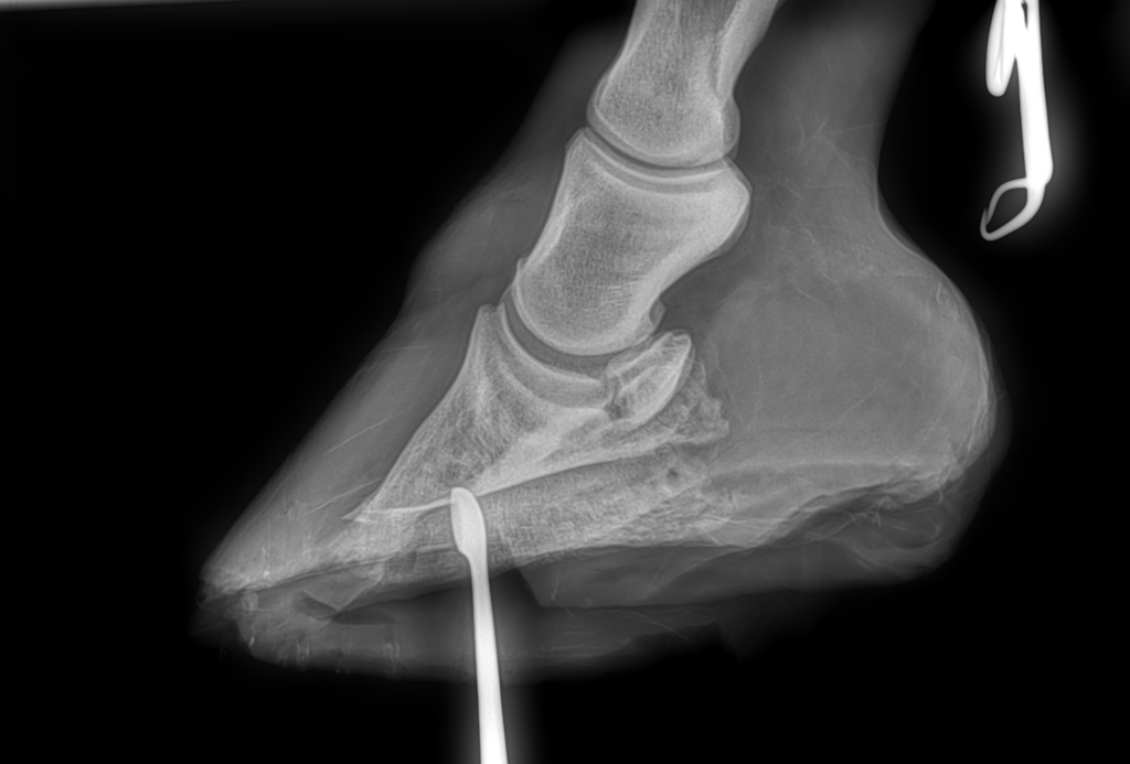
She was diagnosed with infectious pedal osteitis in the left fore and early stages of support limb laminitis in the right fore. Pedal osteitis is an infection of the coffin bone. Annabelle previously showed in the Adult Jumpers with her owner, Jennifer Knobel, but the infection had advanced to the point that she could barely walk.
Local veterinarian Dr. Kim Snyder vigorously managed Anabelle’s foot condition in the field before referring the case to Palm Beach Equine Clinic. She performed digital radiographs at the farm, but they failed to adequately define the lesion. She requested an MRI study of the foot to more accurately image the underlying causes of the persistent foot abscess, such as a foreign body, bone infection, sequestrum, or tumor.
Advanced Medical Imaging Technology
 As the radiographs were not definitive enough alone, the team at Palm Beach Equine Clinic used their state-of-the-art MRI technology for a more detailed analysis. The MRI showed excess fluid and severe inflammation within the coffin bone. The infectious pedal osteitis had caused the bottom of the coffin bone to begin to deteriorate. Worsening the situation, Annabelle was reluctant to bear weight on her left foot. Then she developed mild laminitis in her right foot due to the increased load on the supporting limb to relieve pressure. The development of support limb laminitis determined the emergency status of the left fore lameness as laminitis could be a fatal complication.
Dr. Davis and the team at Palm Beach Equine Clinic took immediate action using the MRI scans for accurate surgical mapping. The MRI study documented exactly where the infectious pedal osteitis was located. This mapped position was used in comparison to the previously taken radiographs. Both imaging modalities were used intra-operatively to guide Dr. Davis precisely to the area of infected bone.
Surgery and Recovery
Annabelle was placed under general anesthesia, and the left front limb was prepared for surgery. A 5 cm long x 3 cm wide window was cut through the sole down to the surface of the coffin bone, through which the necrotic (infected) bony tissue was removed. Prior to recovery, the surgical site was packed with antibiotic powder and a full distal limb bandage was applied. Additional support of Soft-Ride boots and sole supports were positioned. Medical grade maggots were applied to the foot the day after surgery to safeguard absolute full debridement of all necrotic/infected tissues.
 Annabelle was placed on a range of antibiotics. Pain was managed with local nerve blocks and anti-inflammatory medications. Her pain improved rapidly in the operated left limb and signs of laminitis in the right fore resolved as well. During her stall rest recovery, the foot was soaked routinely in Epsom salts. Betadine soaked gauze was applied over the frog and surgery site each day and the hoof was wrapped for protection.
Snyder continued to care for Annabelle in the post-op period. Synder’s husband and farrier, Jim Gilchrist, applied a shoe with a treatment plate to protect the bottom of the foot. This allowed for routine access by removal of three simple screws.
The initial cause of the abscess or why the infectious pedal osteitis progressed so aggressively is still unclear. Dr. Davis speculates that it was possibly a spontaneous abscess and for whatever reason, whether the depth into the foot or the presence of a highly infectious bacteria, Annabelle was just unlucky. Instead of draining like a “normal” abscess, this particular case established an infection within the bone. This made it impossible to clear the infection with even the most aggressive medical therapies apart from surgical removal.
 Annabelle’s infectious pedal osteitis has cleared and her hoof is healing well. Now she is walking sound and has a favorable prognosis for further improvement with hopes of eventually return to work.