Month: September 2017
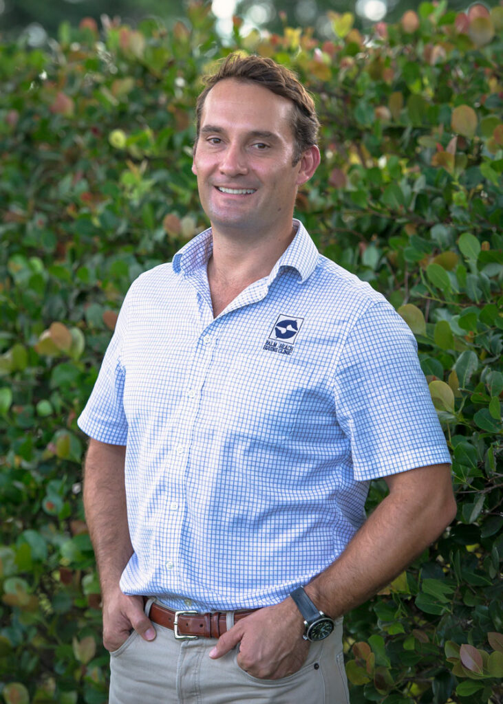
Meet Surgical Resident Dr. Michael Myhre
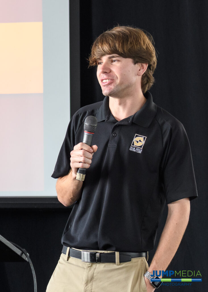
Dr. Michael Myhre was born to be a veterinarian. In 1978 his father, Dr. Grant Myhre, developed a referral practice, Myhre Equine Clinic in Rochester, NH. After working alongside his father since middle school, Dr. Myhre, who hails from Milton, NH, believes he was always destined to be a veterinarian. Dr. Myhre graduated from Cornell University College of Veterinary Medicine based in Ithaca, NY, in 2016, and he joined Palm Beach Equine Clinic thereafter as a surgical resident to work under the direction of board-certifed surgeons Dr. Robert Brusie, Dr. Jorge Gomez, and Dr. Weston Davis.
What is your background with horses?
I grew up in my father’s practice. He would bring me along to see outpatients and cut colics at 2 a.m. When I was in high school and college, I would work there during the summers as a technician. I kept learning from him and when it was time to decide what I would do, I applied to vet school.
We had some lesson horses at home and taught some therapeutic riding, so I rode on the trails occasionally, but I knew I was always supposed to be a veterinarian.
Where did you complete your undergraduate degree?
I attended Ithaca College in New York and studied computer science. It is a pretty unusual undergraduate degree for a veterinarian, but I did not want to go the traditional route of getting a biology degree. Computer technology is now involved in a lot of veterinary medicine – so much of what we do is going through computers, so it is an asset to have that degree.

I still took all the biology and chemistry classes at the same time, and I finished in three years. At that point, I applied to Cornell University and was accepted.
What led you to Palm Beach Equine Clinic?
I came here because it is the best residency program in the country. I have a big caseload and get to work on the best horses in the world. I started on July 1 and what I like the most is the diversity in cases. We have seen hunters, jumpers, dressage horses, and racehorses. I have done everything from condylar fracture repairs to MRIs, nuclear scintigraphy, x-rays, and even colic surgery on a miniature horse. Palm Beach Equine Clinic stays at the forefront of technology with a new standing surgery pit, standing MRI machine, and paperless medical records.
What goals do you have for your veterinary career?
After my three-year residency at Palm Beach Equine Clinic, I plan to move back to New Hampshire and work at my father’s practice.
What can we find you doing when you’re not working?
I am pretty much always working, but my girlfriend is a neurology resident in Manhattan, so I try to visit her as much as I can, or I take advantage of living in Florida and go swimming.
Name one thing most people wouldn’t know about you?
I rowed for the Ithaca College crew team and while I was in vet school, I was an assistant coach for the Cornell University team.
Success Story: Freeman
In January 2016, the Pine Hollow team noticed something seemed off just before driving out of the Winter Equestrian Festival (WEF) with their horses. Stopping to check the horses before continuing off the showgrounds, Pine Hollow discovered Freeman, a promising and successful Dutch Warmblood, had swung his hind leg over the back of the trailer. Freeman’s stifle had ended up squarely on one of the hooks used to secure the back door, lodging the hook into his stifle and into the femoropatellar joint.
Emergency Veterinary Care
Recognizing the extreme peril facing Freeman, Pine Hollow immediately called for help from Palm Beach Equine Clinic, the Official Veterinarians of WEF.
“It took tremendous effort, creative thinking, and exceptional teamwork to free Freeman from the hook impaling his leg,” said David Blake, Pine Hollow’s internationally acclaimed rider and trainer. “Palm Beach Equine Clinic sent several of their top vets to help us rescue Freeman. The team of vets is truly great.”
Thanks in very large part to the help and determination of the vets, Pine Hollow and Palm Beach Equine Clinic were able to free Freeman from the trailer door.
At the Equine Hospital
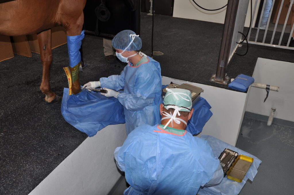
From there, Freeman was transported to the nearby Equine Hospital, where he spent a few days recovering before it was agreed to pursue arthroscopic surgery on his femoropatellar joint.
“To be honest, it wasn’t looking good at all for the first day or so Freeman was there,” said Blake. “The joint was so severely damaged we didn’t know if it could be fixed. Our only chance of fixing the joint was surgery, so we agreed we would try everything possible.”

Dr. Weston Davis performed the surgery, after which Freeman remained in Palm Beach Equine Clinic’s care while he regained use of the leg.
“The team did a fantastic job there and kept Freeman until he was ready to begin long-term rehab with James Keogh,” said Blake.
When Freeman was finally ready to return home to Pine Hollow, Blake hoped at best Freeman would eventually be able to do light work and perform at a low level.
In the past, when a horse’s gait has felt off or lacking in its usual impulsion, it was often assumed to be an issue of lameness. Now however, thanks to the improved diagnostics readily available at the Palm Beach Equine Clinic veterinarians are able to more accurately pinpoint the problem area. Perhaps surprisingly, it’s not always in the legs or hooves. With increasing frequency, the horse’s neck is being diagnosed as the root of the issue.
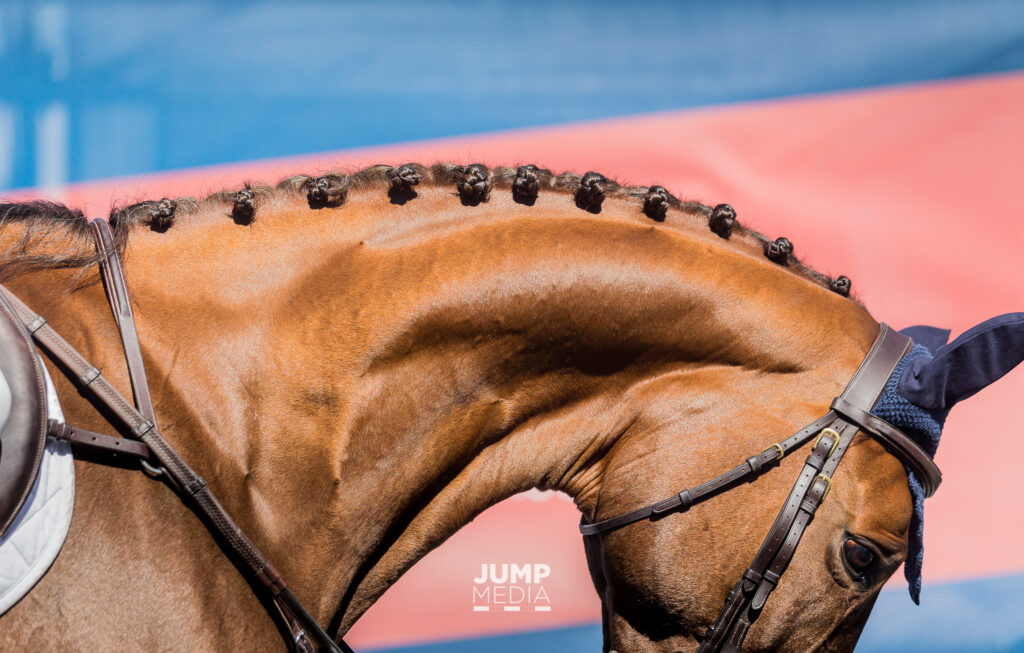
The Anatomy of the Equine Neck and What Can Go Wrong
In order to understand the problems that can arise in association with the horse’s neck, it’s important to first understand the anatomy.
The neck is composed of seven cervical vertebrae running from the head to the thorax, named C1 through C7, and each articulating with each other. The primary purposes of the neck are to move the head and to protect and transport the spinal cord and nerves, which run through the middle of the vertebrae.
Such a major role as the protection of the nerves and spinal cord can also come with some major risks and complications, with clinical signs of these problems generally presenting themselves either neurologically, as neck pain, or as lameness in the front legs. These more specific symptoms may include:
- Ataxia or clumsiness – Ataxia is defined as the “lack of control of bodily movements”. In the case of an ataxic horse, you may begin to notice staggering, sudden loss of balance, or even an inability to remain upright. Ataxia is generally an indicator of a neurological condition or damage to the spinal cord itself, caused by either developmental issues, trauma, or an infectious disease such as equine protozoal myeloencephalitis (EPM). Such neurological cases can often be the most debilitating.
- Lameness – You can think of the spinal cord and the nerves in the neck like an interstate, with the spinal cord itself acting as the major highway. As you are “driving” along the interstate, every so often there are little exits, which is where the other nerves come out. Should there be any impingement on the interstate or spinal cord itself, you’re likely looking at more severe complications – much like an accident on the highway. Should there be impingement on the nerves coming off of the spinal cord, it will more likely present itself like an accident a little way off an exit – not affecting the interstate itself, but possibly causing problems that spread elsewhere. That is where we see lameness issues arise.
This can be more difficult to pin down, but can often be due to pressure on the smaller nerves that pass through the openings in the vertebrae and supply the front legs. Arthritis of the articular facet joints of the vertebrae is another common reason for lameness, as anytime these joints become arthritic or inflamed, it can easily translate to the forelimbs.
- Neck Pain – This will often go hand-in-hand with lameness, as factors such as arthritis of the articular facet joints can lead to both symptoms. Other possible reasons for neck pain include trauma or inflammatory diseases.
Diagnosing the Problem
Neck problems, particularly those related to lameness, are generally diagnosed through a process of exclusion, first performing nerve blocks to or ruling out lower regions of the horse’s body. Palpation of the neck, testing of the neck’s movement, and full neurological exams may also be performed in addition to a full lameness exam, depending on the horse’s symptoms.
Once other regions of the horse are ruled out as the location of the problem, veterinarians are now able to use diagnostic images such as radiographs, nuclear scintigraphy and standing CT scans to specifically locate problems in the neck like never before.
In years past, those diagnostic resources were left for last-ditch cases when veterinarians really could not pinpoint any other problems. Today, with the advent of more modern technology, better radiographs, better ultrasound machines, and the more advanced imaging of nuclear scintigraphy and CT scans, veterinarians are able to readily utilize advanced diagnostics to save time and money, and to find the root of the problem more quickly and accurately.
The neck is one of the areas that has most clearly benefited from the progression in advanced imaging. While neck problems have likely been prevalent for some time, veterinarians are now finding those diagnoses more common, as they are able to more accurately locate the issue – particularly at clinics like Palm Beach Equine Clinic. Often these issues will go undiscovered or undiagnosed in the field, but they can be identified at Palm Beach Equine Clinic thanks to the readily available imaging tools.
As an example, if arthritic factors are suspected, nuclear scintigraphy can be used to look for areas of increased bone turnover. On the resulting bone scan, veterinarians are looking for areas where there is an increased calcium uptake because the bone is actively remodeling. These areas will appear darker on the scan and are generally a good indicator of a boney injury or arthritis, with the darker “hot spots” often appearing above the articular facet joints.
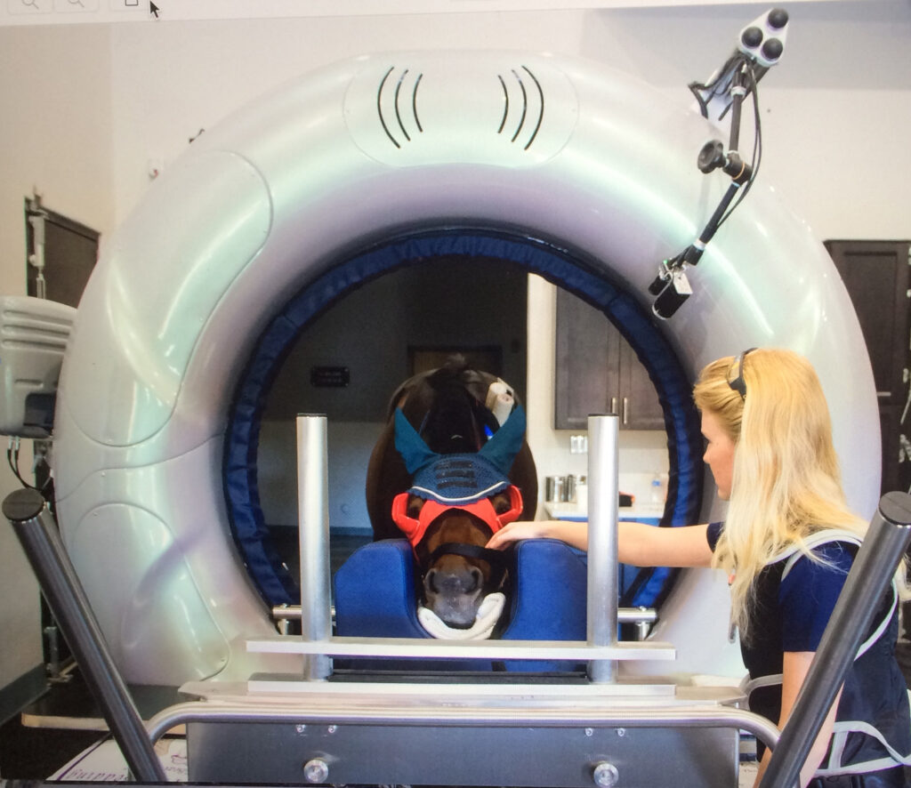
Also new and groundbreaking for the diagnosis of equine neck problems is the use of a Computerized Tomography or CT scan, with the ability to scan a standing horse with light sedation on the near horizon which will be available at the clinic this upcoming winter season.
Treatment
Once a solid diagnosis is arrived upon, the proper treatment protocols can be prescribed. Depending on the root of the problem, possible treatments may include shockwave therapy, regenerative therapies such as interleukin-1 receptor antagonist protein (IRAP) therapy or platelet-rich plasma (PRP) therapy, or one of the most common treatments, injections of the facet joints.
In the case of facet joint injections, veterinarians at Palm Beach Equine Clinic are able to medicate under ultrasound guidance, guiding a needle into the joints and delivering corticosteroids or similar medication. Surgery is also an option as a final approach to severe complications.
In milder cases, treatments may also just call for increased time off, chiropractic treatments, or the administration of non-steroidal anti-inflammatory (NSAID) medications.
If you suspect any issues with your horse’s neck, contact Palm Beach Equine Clinic any time by calling (561) 793-1599 to schedule an appointment.