Tag: computed tomography
Typically, when a horse’s gait feels off or may be lacking usual impulsion, the rider often assumes it to be an issue of lameness associated with the forelimbs or hindlimbs. However, that may not always be the case. Utilizing advanced diagnostic imaging techniques, Palm Beach Equine Clinic is able to accurately pinpoint the specific area that is affecting overall performance. In many cases, the cervical vertebrae are often identified as the cause of lameness, asymmetry, and poor performance.
Vertebral Anatomy

The neck is composed of seven articulating cervical vertebrae running from the head to the thorax, named C1 through C7. The neck allows movement of the head while protecting the spinal cord and providing an avenue for nerves to travel. Impingement on the spinal cord and nerves connected to the cervical vertebrae can exhibit neurologically as ataxia, neck pain, or lameness.
Signs of Lameness Related to the Neck
In a lameness exam, a veterinarian will perform flexion tests and palpate areas of the body looking for decreases in the horse’s range of motion or pain upon flexion. The rider may pick up on subtle lameness issues associated with the neck by feeling a change in the horse’s suppleness or resistance to yielding in a certain direction. Lameness may even present itself as a difference in the horse’s balance, such as being heavier on the forehand, or performance issues such as late lead changes. The tried-and-true “carrot test” can also show if a horse is resistant to flexing their neck.
Identifying Lameness through Diagnostic Imaging
Historically, neck issues related to performance are generally diagnosed through a process of ruling out other areas of the body. Diagnostic imaging can now be the most powerful and effective tool for identifying the cause of lameness related to cervical injury and hereditary malformation.
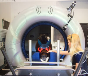
Computed Tomography (CT) scans have revolutionized the ability to assess the entire neck and can be performed while the horse is standing and under light sedation. Computed Tomography images can be rendered into three-dimensional models and sliced in any orientation, allowing the veterinarian to evaluate the vertebrae in great detail that is incomparable to standard radiographs (x-rays). These comprehensive CT scans offer veterinarians a thorough profile so they can accurately diagnose and initiate an effective response.
A standing CT scanner is the latest addition to Palm Beach Equine Clinic’s arsenal of diagnostic imaging modalities. Currently, Palm Beach Equine Clinic is the only equine hospital in South Florida offering this capability. Compared to other modalities such as MRI or Nuclear Scintigraphy, Computed Tomography offers a valuable return for its rapid acquisition of images. If you suspect there is an issue in your horse’s neck please, contact Becky at Palm Beach Equine Clinic at 561-793-1599 to schedule an appointment.