Tag: weston davis
Success Story: Freeman
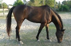
In January 2016, the Pine Hollow team noticed something seemed off just before driving out of the Winter Equestrian Festival (WEF) with their horses. Stopping to check the horses before continuing off the showgrounds, Pine Hollow discovered Freeman, a promising and successful Dutch Warmblood, had swung his hind leg over the back of the trailer. Freeman’s stifle had ended up squarely on one of the hooks used to secure the back door, lodging the hook into his stifle and into the femoropatellar joint.
Emergency Veterinary Care
Recognizing the extreme peril facing Freeman, Pine Hollow immediately called for help from Palm Beach Equine Clinic, the Official Veterinarians of WEF.
“It took tremendous effort, creative thinking, and exceptional teamwork to free Freeman from the hook impaling his leg,” said David Blake, Pine Hollow’s internationally acclaimed rider and trainer. “Palm Beach Equine Clinic sent several of their top vets to help us rescue Freeman. The team of vets is truly great.”
Thanks in very large part to the help and determination of the vets, Pine Hollow and Palm Beach Equine Clinic were able to free Freeman from the trailer door.
At the Equine Hospital
From there, Freeman was transported to the nearby Equine Hospital, where he spent a few days recovering before it was agreed to pursue arthroscopic surgery on his femoropatellar joint.
“To be honest, it wasn’t looking good at all for the first day or so Freeman was there,” said Blake. “The joint was so severely damaged we didn’t know if it could be fixed. Our only chance of fixing the joint was surgery, so we agreed we would try everything possible.”

Dr. Weston Davis performed the surgery, after which Freeman remained in Palm Beach Equine Clinic’s care while he regained use of the leg.
“The team did a fantastic job there and kept Freeman until he was ready to begin long-term rehab with James Keogh,” said Blake.
When Freeman was finally ready to return home to Pine Hollow, Blake hoped at best Freeman would eventually be able to do light work and perform at a low level.
Shes Packin Fame: Back in Winning Form
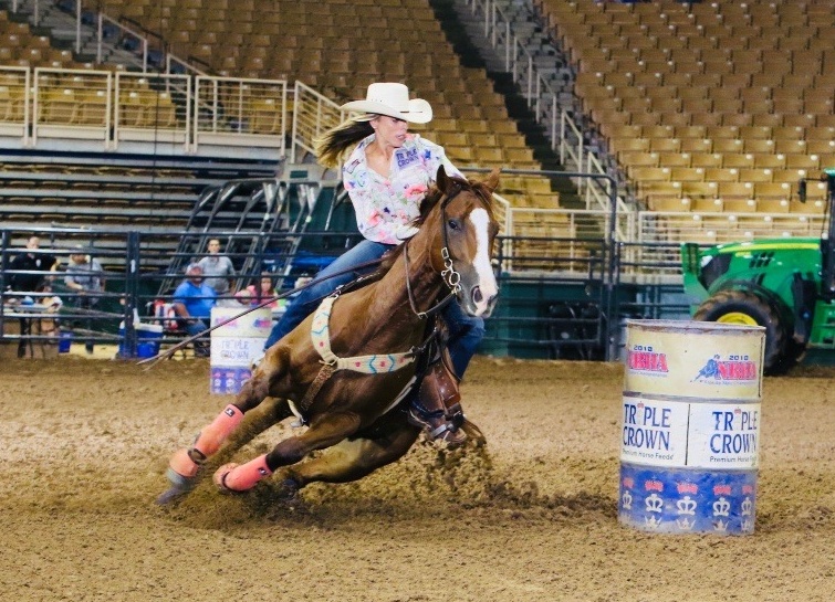
Nearly eight months ago, Shes Packin Fame, a 2012 Quarter Horse mare owned by Margo Crowther of Fort Myers, FL, suffered a rare slab fracture to the central tarsal bone in her left hock while competing in a barrel racing competition. After a diagnosis aided by Palm Beach Equine Clinic’s (PBEC) state-of-the-art diagnostic imaging equipment and a surgery performed by PBEC’s own Dr. Weston Davis, Shes Packin Fame has not only returned to running barrels, the five-year-old mare is back to winning.
Crowther purchased Shes Packin Fame, affectionately known as Sissy, as a three-year-old after the mare reminded her of a horse she ran in college. Crowther trained Sissy herself and won or placed in nearly every barrel futurity she entered during the horse’s four-year-old year, accumulating $100,000 in prize money.
In November of 2016, Crowther and Sissy were competing at the No Bull Finals in Asheville, NC, when Sissy went down at the first barrel on the final day. The fall fractured the horse’s central tarsal bone, which was not easily diagnosed. Crowther met with a veterinarian in North Carolina who was unable to locate the fracture via x-ray before contacting Dr. Davis, who had managed Sissy’s healthcare since she joined Crowther’s string.
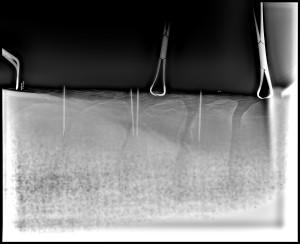
Dr. Davis utilized PBEC’s Equine Standing Magnetic Resonance Imaging (MRI) and Nuclear Scintigraphy (bone scan) modalities to locate a flat piece of separated bone known as a slab fracture.
The process began with a bone scan where Sissy was injected with a radioactive isotope named Technetium 99. The isotope attached to the phosphorous proteins localized within the bone and was absorbed. A specialized nuclear isotope gamma ray camera was used to capture images of the skeletal anatomy with a 360-degree view. Points of interest lit up on the image to indicate increased metabolic activity and was able to locate the site of the injury.
Following the identification of the injured area, a Standing MRI produced highly detailed images in several different planes to capture a compete view of the injury and further define the issue.
After Dr. Davis located and identified the fracture, he surgically inserted a screw into the central tarsal bone to stabilize the fracture. Sissy was discharged from the clinic on six months of recovery with follow-up diagnostic imaging every month to monitor the injury’s repair. During the fourth month of recovery, Dr. Davis removed the screw. At the end of March, Sissy was cleared to begin exercise and Crowther began by hand walking the mare slowly progressing to trotting her under tack. They started with ten minutes of exercise and worked up to 45 minutes.
“Weston was a huge part of Sissy’s recovery,” said Crowther, who set her sights on entering Sissy in the Old Fort Days Derby, held over Memorial Weekend in Fort Smith, AR. “It is the biggest derby of the year for five-year-olds. When it came time to enter, Weston rechecked the leg, did flexion tests, cleared her to run, and wished me good luck.”
When they arrived in Fort Smith, Sissy had not seen a barrel since the day of the injury. Crowther and Sissy posted a time of 16.405 seconds, the fastest time of the event, to win the 25-horse final and collect a $23,469 prize money check.
“She just came back so confident and so strong, like she never missed a beat,” said Crowther. “She always ran like an older horse, but I was surprised at her time. I knew she would be in the top ten, but I was surprised just how strong she was. Weston told me to let her set her own pace and that is what I did. I did not push her. So, when I called Weston to tell him we had won, he was very surprised.
“She feels like her hock is maybe even stronger than it was before the injury,” continued Crowther. “I am so thankful to Weston and Palm Beach Equine Clinic, and feel blessed that she has come back strong and healthy.”
With Sissy back in top form, Crowther’s next goal is a lofty one. Her hope is to qualify for and compete at the National Finals Rodeo in Las Vegas, NV, this December.
At Palm Beach Equine Clinic in Wellington, FL, the team of Board-Certified surgeons are experts in minimally invasive surgical techniques, aiming to reduce joint disease, resolve lameness, and improve the longevity of sport horse careers.
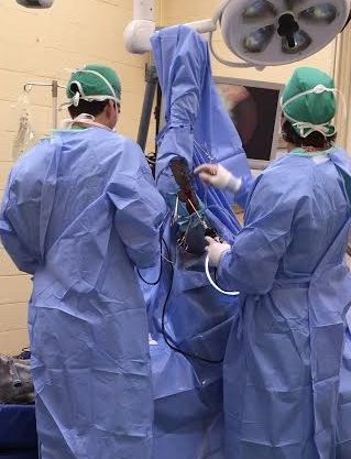
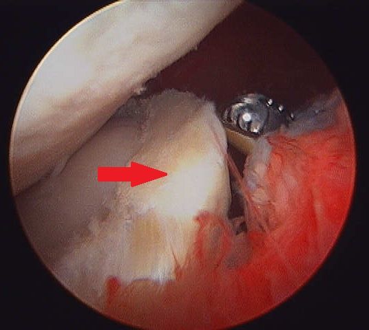
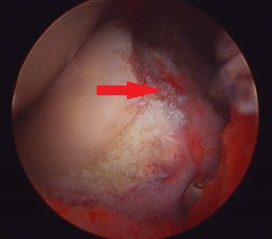
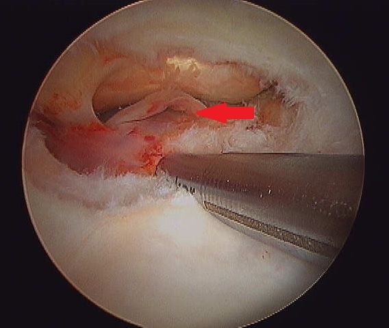
Arthroscopy (or arthroscopic surgery) is a minimally invasive surgical technique that can be performed on an injured joint or synovial structure to accurately explore and treat pathology. The surgery generally involves two very small (8mm) keyhole incisions. The first incision is where the surgeon will insert the arthroscope, which is an instrument with a small surgical grade camera installed that allows a complete, clear view of the interior joint surface. The second small incision is created to insert the surgical instrument to perform the procedure.
Arthroscopy is used to treat a broad range of injuries inside of a joint. Chip fracture removal is a procedure that is particularly commonly in both young Warmblood horses with developmental disease and in racehorses travelling at high speeds. A small chip fracture can cause persistent irritation in the joint as well as arthritis if left untreated. It is best removed immediately so that no further damage is created. The surgeon can go into the joint, remove the chip, and clean up the surrounding cartilage. Most horses recovery quickly and return to their normal athletic activity.
Board-Certified Surgeon Dr. Weston Davis performs many arthroscopic surgeries at Palm Beach Equine Clinic alongside fellow surgeons Dr. Robert Brusie and Dr. Jorge Gomez.
“In many horses, we consider arthroscopy as a prophylactic measure, intervening after injury, but before the development of a generalized degenerative arthritic cycle ensues,” Dr. Davis stated. “Arthroscopy is definitely something that you want to do early in the game if you feel like the horse has joint disease, or a chip, or cartilage disease, or an undefined injury that is not responding appropriately to medical therapy. Arthroscopy can be curative for some of these horses. But if you do not intervene early on in the course of the disease and there is already advanced arthritis, then you have missed your window.
“Arthroscopy is a preferred treatment because it is minimally invasive so most horses can go right back to work,” Dr. Davis continued. “In a typical scenario, we thoroughly explore the joint with the arthroscopic camera, we remove a chip or repair a lesion, and the horse is not lame after the surgery. Because of the small incisions, there is minimal aftercare and horses are often able to go back to work quickly.”
Other common indications for arthroscopic surgery are meniscal disease in the stifle, subchondral cystic lesions, primary cartilage lesions, and debridement of damaged tendinous/ligamentous tissue (such as deep digital flexor tendon tears in the navicular bursa). The surgeons at Palm Beach Equine Clinic can perform arthroscopy on virtually any joint in the horse. Anything from the Temporomandibular Joint (TMJ) of the head to the navicular bursa within the hoof capsule can be explored and treated with this minimally invasive approach.

Almost all arthroscopies are performed under general anesthesia with the horse on its back. New renovations at Palm Beach Equine Clinic include a set of stocks of adjustable height adjacent to a surgeon’s pit, allowing the surgeons to have eye-level access to the joint they are working on, enabling many new procedures on the legs of standing horses.
Minimally invasive surgery allows for a simple and quick recovery for the horse. The traditional horse would be on stall rest with a bandage on until the sutures come out at two weeks, and then start doing some light hand walking and physical therapy. Barring severe damage in the joint or associated tendon/ligament disruption, most cases will undergo a six-week rest and rehabilitation protocol, then return to normal work.
As always, the advanced diagnostic imaging at Palm Beach Equine Clinic permits the surgeons to get a complete evaluation of an injury involving a joint to ensure the best possible outcome. Depending on the injury type, digital radiographs, ultrasound, MRI, and Nuclear Scintigraphy, or a combination thereof, may be used for pre-operative diagnosis and planning. Ultrasound and digital radiography are available for intra-operative use. Intra-operative CT scanning will also be available in the future with the new additions at Palm Beach Equine Clinic.
“When you are inside the joint with an arthroscopic camera, you have the most complete picture of the surface and health of that joint,” Dr. Davis noted.
Palm Beach Equine Clinic of Wellington, FL, is a worldwide leader in sport horse medicine and emergency colic care. While symptoms of colic should be treated medically first, surgical intervention can be necessary, and the team at Palm Beach Equine Clinic is prepared for every situation.
With three Board-Certified Surgeons on staff, as well as a state-of-the-art hospital and the most advanced surgical equipment, Palm Beach Equine Clinic has a very high success rate in saving horses from life-threatening colic. The veterinarians take pride in their equine clients returning to full intended use and continuing to perform at their highest levels.
Causes and Symptoms
Colic is defined as any source of abdominal discomfort in the horse. Abdominal pain or problems within the gastrointestinal tract can arise unexpectedly from many different origins, including but not limited to: spoiled feed, abrupt changes in feed, parasite infestation, sand ingestion, lack of water consumption, excess stress, or changes in the weather. Many times there is not a well-defined inciting cause.
The most important step any owner can take is to recognize the symptoms as early as possible and immediately call their veterinarian. Pawing, rolling, looking at the abdomen, sweating, loss of interest in food and water, and absence of gut sounds in any of the four quadrants are common symptoms. The sooner the veterinarian gets involved in treatment, the better the horse’s chance of survival.
In the event of an emergency, the surgeons and veterinarians of Palm Beach Equine Clinic are available 24/7. When an equine patient is admitted to the hospital, every step is taken to quickly diagnose the problem and correct it immediately.
Tests and Diagnosis
Board-Certified Surgeon Dr. Weston Davis explained that one of the biggest challenges in the sport horse population is determining surgical versus non-surgical colic cases.
“We do not want to put a non-surgical case through the risk of anesthesia and the months of healing time, so we try to spare that at all costs and determine the surgical cases as accurately as we can,” Dr. Davis detailed. “On the split side of that, we try to operate as quickly as possible on any horse that needs surgery and not miss any surgical lesion types.”
There are several methods for differentiating surgical cases. Simple physical exam findings, such as the color of the gums, heart rate, gut sounds, and level of pain can all be supportive of surgical necessity. A variety of tests may also include abdominal ultrasounds and rectal exams.
An Abdominocentesis (or belly tap) is performed on every questionable colic case, where fluid is collected from around the intestines and analyzed for color and character. A variety of other laboratory tests will be run on the fluid as well, with the aim of quickly determining if the horse’s bowel is compromised.
Surgical Procedures
If surgery is necessary, there are a few different approaches that may be performed depending on the specific case.
For chronic colic cases, such horses with longstanding, intermittent colic, an abdominal exploratory procedure may be done with laparoscopy. This option can be done with the horse standing and is a minimally invasive way to examine the full abdomen.
In most acute cases, further steps must be taken. If the veterinarian determines that the horse is a surgical candidate, the patient will go under general anesthesia. The surgeons try to make as small of an incision as they can to perform the needed surgical correction.
“If we intervene early, we can take a strangulating or compromised lesion – one that most people understand as a twist – and we can go into the abdomen and correct the twist, reposition everything appropriately, explore the remainder of the abdomen to make sure nothing else is going on, and then close them up,” Dr. Davis explained. “Some of these surgeries can be as quick as 30 minutes and require just an untwisting, repositioning, and closure. The ones that are bad are the usually the cases that have a more severe twist or have been going on longer.”
In more severe or long-standing cases, the surgery can require a resection and an anastomosis procedure to excise a compromised or devitalized segment of the intestine. The surgeon then joins the healthy ends back together.
“Even more advanced procedures would be like a re-plumbing of the intestines,” Dr. Davis noted. “One of the most common examples of this would be a patient with damage to the end of the small intestine, near or involving its junction with the cecum. In a case like this, we would perform a ‘jejunocestomy’ where we join another part of the small intestine to a different position on the cecum.”
Post-Operative Recovery
After any surgery, there is a process of recovery, which Palm Beach Equine Clinic makes as easy as possible for its clients. In the traditional recovery, most horses will remain in the hospital for a few days. In the post-operative period, they generally receive fluids until they are ready to eat and drink, 3-5 days of antibiotics, and 5-7 days of anti-inflammatories.
“The recovery process is highly dependent upon how sick they are after surgery,” Dr. Davis stated. “Some horses will bounce back and be home 48 hours later, but a very sick horse could potentially spend seven to ten days in the hospital until they are healthy enough to get off fluids and go home.”
After leaving the hospital, the horse is usually placed on one month of stall rest, followed by another month of turnout in a small paddock. In between eight to 12 weeks, the horse will usually be fully recovered and ready to start back to work.
Physical Therapy
With the sport horse in mind, PBEC pays special attention to the health of the abdomen following surgery. The health of the abdominal incision and prevention of infection or hernias is very important. In most cases, the surgeon will recommend physical therapy and special exercises to re-strengthen the horse’s abdominal muscles so that it can get back to work quickly and have a strong abdominal musculature when it does.
“Making the horse walk backwards is one thing that will make them tighten and work their abdominal musculature,” Dr. Davis shared. “Pinching or tickling around their tail head is another common exercise to make them do something similar to a stomach crunch.”
As one of the top equine emergency care centers in the world, Palm Beach Equine Clinic is prepared to handle any case, 24 hours a day, seven days a week, and 365 days a year.
“With the combination of quick surgical intervention, excellent surgical care, and specialized post-operative measures, Palm Beach Equine Clinic has a very high rate of return to athletic performance for all of our colic cases,” Dr. Davis concluded.
Palm Beach Equine Clinic provides experience, knowledge, availability, and the very best care for its clients. To find out more, please visit www.equineclinic.com or call 561-793-1599.
Learn More About Surgeon Dr. Weston Davis

Dr. Weston Davis is a second-generation veterinarian from South Florida. His father is recently retired from veterinary medicine and his family raises beef cattle in Clewiston, FL. Dr. Davis graduated Magna Cum Laude from the University of Florida College Of Veterinary Medicine in 2008. He was awarded the Barbaro Gulfstream Scholarship (a veterinary scholarship named in honor of the amazing Barbaro), the Calder Race Course Scholarship, and the Student Award for Excellence in Large Animal Surgery.
After graduation, Dr. Davis completed his internship in Sports Medicine and Surgery at Oakridge Equine Hospital, followed by a residency in Equine Surgery at North Carolina State University. In 2012, he became board certified in Large Animal Surgery by the American College of Veterinary Surgeons. Before joining Palm Beach Equine Clinic, Dr. Davis spent 1.5 years as a staff surgeon at a private practice referral center in Texas. He has authored and co-authored publications on topics ranging from colic surgery to advanced imaging and novel surgical techniques. In 2014, he was awarded the BEVA Trust Peter Rossdale EVJ Open Award for a research publication on return to performance following colic surgery. He has spoken at several national and state meetings.
Dr. Davis is an avid sportsman himself, and his hobbies include fishing, hunting, waterskiing and almost any outdoor activity.
Tell us more about your background with horses growing up?
I started riding horses when I was so young I can’t remember. We had some amazingly kind horses that packed us around and took care of us (and a couple that didn’t). Riding as a child was mostly business – for the purpose of working cows. Somewhere around 14, I began team roping for pleasure and competition. I roped throughout college, but lost the required free time when I began practicing veterinary medicine. I’ll probably get after it again when I am retired and much too old to be doing that sort of thing!
When and why did you decide to become a veterinarian and why did you choose to pursue a career in surgery?
I decided to be a veterinarian very early in life.I had a father and uncle who were both successful and happy veterinarians whom I looked up to, so it was a logical path to follow. The surgical interests started as a kid watching my father do surgery, something I always thought was amazing and he was very skilled at. My decision to pursue the surgical avenue came during vet school when I realized that I wanted to specialize and knew surgery was my passion.
What is the best advice that your father has given you as a veterinarian?
My father is a man of few words. However, by watching him, I learned one of the biggest life lessons, which is to be calm and content with your career and your life.
What do you enjoy about speaking publicly and sharing your knowledge?
I think mentoring, sharing knowledge and teaching the hands-on skills to the next generation of veterinarians is one of the most fun and rewarding parts of my job. Observing a student as content and excelling in their career with a skill set that you contributed to, even in a small way, is a beautiful thing.
What is the most interesting or rewarding surgical case you have worked on?
Although minimally invasive arthroscopic type surgeries are my favorite to perform, I think colic surgeries are one of the most rewarding. They are often difficult surgeries and inevitably in the middle of the night, but you take a horse who would most certainly die without you, and save a life. Helping a horse who was in excruciating pain or has a life-threatening devitalized piece of intestine recover back to feeling comfortable and eating in their stall the next day is about as good as it gets.
What are your goals for your career now?
My goal is to expand the surgical and outpatient sports medicine referrals at PBEC. I am currently the coordinator for the intern and resident program. I plan to expand and improve on the quality of these mentoring programs within the industry. I am also working on the development and description of some novel minimally invasive surgical techniques. I aim to continue authoring 1-2 publications annually in the refereed equine literature.
When not roping, fishing, hunting or water-skiing, what other things do you do with your free time?
Most of my non-equine time lately has been devoted to some real-estate interests and house renovations. I am also a big reader.
Jo Ann Hopkins and her 12-year-old Appendix-bred gelding, Blazer, were placed third in points in their local 1D barrel racing circuit. However, three months earlier, Blazer underwent surgery at Palm Beach Equine Clinic for a growing cyst in his sinus. On August 1, 2015, Dr. Weston Davis, DACVS, performed the surgical procedure on Blazer for an expansile paranasal sinus cyst of the left paranasal sinuses, extending to the right concho-frontal sinus.
Hopkins noticed that Blazer, who she calls “George” for his laid-back attitude, had a raised knot on his head this spring and consulted with her local vet, Dr. Kelly Alderman. The area was monitored closely and remained in a similar condition until June, when Hopkins noticed Blazer wheezing while running barrels and the knot increasing in size.
Diagnosing the Paranasal Sinus Cyst
Dr. Alderman performed radiographs on the site and aspirated the swelling where a substantial amount of fluid was removed. She confirmed that the bump was a suspected paranasal sinus cyst. At that point, Blazer was referred to Dr. Weston Davis of Palm Beach Equine Clinic, who advised Hopkins that the cyst was likely growing slowly within the sinus for a considerable amount of time with no clinical signs. The diagnosis was an expansile paranasal sinus cyst of the left paranasal sinuses, extending to the right frontal sinus.
Paranasal Sinus Cyst Surgery
For surgery, Blazer was placed under standing sedation and local analgesia (painkiller) and his head was aseptically prepared before Dr. Davis performed a frontonasal sinusotomy (incision into the sinus). He made an opening from the middle of Blazer’s head to the corner of his eye. Once the skin flap was elevated and the bone fractured, a large amount of fluid was evacuated, which is consistent with the contents of a paranasal sinus cyst. Dr. Davis also observed that the sinus cavity was extremely distorted and expanded. The cyst lining was debrided and removed, then the sinus was lavaged and a tube was advanced into the sinus cavity to facilitate drainage. The sinus was packed with gauze tied together and soaked in a dilute betadine. Finally, the bone flap was replaced and the skin was closed and covered with a sterile bandage.
Recovery
The gauze packing was removed 48 hours after surgery and upon release from Palm Beach Equine Clinic, Hopkins was instructed to monitor the incision for heat, swelling and discharge. Bute, in the amount of 2g, was administered orally once a day for five days and Dr. Alderman removed the sutures after 10 days.
Hopkins characterizes Blazer as a sweet gelding with heart to spare, and is happy to report that his recovery has been fantastic. After six weeks of stall rest and rehab time to get back in shape, Blazer recently won his first barrel race after surgery. According to Hopkins, Blazer is running better than ever thanks to Dr. Davis and the staff at Palm Beach Equine Clinic.
Palm Beach Equine Clinic is renowned for its exceptional care of performance sport horses of all disciplines around the world. Sometimes, the veterinarians and surgeons have the opportunity to treat the horses owned by their colleagues. This summer, Palm Beach Equine Clinic veterinary technician Megan O’Neal experienced the clinic’s surgical expertise firsthand when her rescued off the track Thoroughbred (OTTB) was diagnosed with severe spinal impingement (kissing spines).
Signs and Symptoms
O’Neal adopted Blessing, a 10-year-old mare, almost three years ago. After Blessing’s career on the track ended, she was given to Pure Thoughts Horse Rescue in Wellington, FL. Blessing had moved through a few foster homes before O’Neal gave her a forever home. Blessing had been successfully jumping three-foot courses with one of her previous foster homes. After adopting Blessing, O’Neal was jumping the mare as well, but only about two-feet high. She had plans to compete, but Blessing’s behavior changed under saddle. The mare began rearing and bolting, endangering both herself and her rider. Her attitude on ground handling turned sour as well, no longer enjoying being groomed and pinning her ears in agitation.
Identifying the Problem

With the help of Palm Beach Equine Clinic surgeon Dr. Weston Davis and the advanced imaging technology available at PBEC, O’Neal was able to pinpoint the cause of Blessing’s troubling change in behavior. The diagnosis was severe chronic back pain with dorsal spinous process impingement (kissing spine lesions). The vertebrae in her back from T16 – L1 were affected, which is the mid-section of the horse’s back, in the general region of where the back of the saddle sits.
Addressing the Problem Through Non-Surgical Treatments
The first course of action was to try several non-surgical techniques to treat Blessing’s pain. Dr. Davis tried treatments of intramuscular injections for arthritic pain and corticosteroid injections in between the spinal vertebras. A veterinary chiropractor adjusted the mare every few weeks. For almost eight months, several additional methods were tried to avoid kissing spines surgery including acupuncture, back stretches and oral muscle relaxants. In July, when the pain continued after all their efforts, O’Neal finally decided that surgery was the only option for recovery.
Kissing Spines Surgery
In the state-of-the-art hospital at Palm Beach Equine Clinic, Blessing was sedated to obtain pre-operative radiographs that map the exact site of the lesions. She was placed under general anesthesia and the surgical site was sterilely prepared. Dr. Davis made a single 20cm incision on the dorsal midline over the palpable dorsal spinous processes. Eighteen-gauge needles were inserted at regular intervals and used as radiographic markers to identify the interspinous spaces. The incision was extended through the supraspinous ligament at each site.
Dr. Davis used sterile surgical equipment, known as a bone rongeur, to elevate the soft tissues and resect some of the affected dorsal spinous processes (DSPs). The rongeurs, which are similar to large pliers, were used to slowly remove the edges of the overriding bone. This process frees the space between adjacent vertebrae until widened enough that the index finger of the surgeon could easily pass in the interspinous space. The site of resection was lavaged to remove any loosed tissues. Lastly, Intra-operative radiography was used as needed to confirm the location and completion of the surgery. Following confirmation, the supraspinous ligament was internally closed with absorbable sutures. The skin was closed with surgical staples and a stent was sutured in place over the incision. After several weeks of healing, the stent and staples were removed.
The kissing spines surgery was performed without complication and Blessing was provided a good prognosis. Soon after surgery, the space that was created between vertebrae filled with a non-painful, fibrous scar tissue. Blessing went home the following day after surgery. She was monitored closely at home and received routine peri-operative antibiotics (gentamicin and penicillin) and anti-inflammatories (phenylbutazone) for pain. Recently, O’Neal was given the okay from Dr. Davis to resume riding for normal exercise. O’Neal looks forward to continuing her partnership with this special mare. Thanks to Dr. Weston Davis and the team at Palm Beach Equine Clinic for their exceptional care to get Blessing back to happy and healthy!
Thoroughbred Brazilian Triple Crown winner Bal a Bali was admitted to Palm Beach Equine Clinic on August 3, 2014. The elite athlete was treated for life-threatening laminitis by board-certified surgeon Dr. Weston Davis of Palm Beach Equine Clinic, in conjunction with Dr. Vernon Dryden, just months after his Triple Crown win in March of that year.
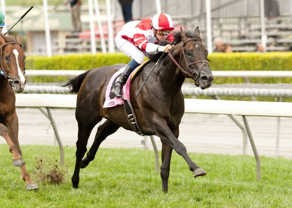
Brazil’s 2014 Horse of the Year, Bal a Bali (Put It Back—In My Side, by Clackson) took an impressive win in the Grande Premio Cruzeiro do Sul (Brz-I) to become the country’s 12th Triple Crown winner. He finished the race in track-record time at Gavea racecourse.
Following his last start in June 2014, Bal a Bali was purchased by Fox Hill Farm and Siena Farm and imported to the U.S. in late summer, but unfortunately suffered from laminitis brought on during his travels. Bal a Bali was in a Florida quarantine center scheduled to fly to trainer Richard Mandella’s stable in California when the problems developed.
Bal a Bali Admitted to PBEC Equine Hospital
Bal a Bali was quickly moved to Palm Beach Equine Clinic in Wellington, Florida, where he was received by Dr. Weston Davis, who would oversee his care in the equine hospital for the next three months. Palm Beach Equine Clinic set aside an entire section of the hospital barn as a quarantine unit to meet the horse’s final import requirements while he was treated with aggressive cryotherapy – a gold standard of laminitis care. Hospital staff carefully monitored Bal a Bali and treated him with consistent cold-water spa treatments for several days throughout the severe acute phase of this disease. He was gradually weaned out of the spa as he improved clinically.
On two occasions, Dr. Davis performed intravenous regional perfusions of the horse’s feet with advanced stem cell treatments. A myriad of other medical therapies were administered throughout his stay. The progression of his laminitis was closely monitored with the use of diagnostic imaging and meticulous farrier care. Farriery care included ensuring optimal sole support and proper mechanics to decrease strain on the fragile lamina. By October, the horse was cleared to travel to Siena Farm in Kentucky. There, Dr. Dryden continued to treat the horse and he was then flown to California in January.
Winning his Battle with Laminitis
After a nine-month recovery process, Bal a Bali made a miraculous return to the track for his North American debut in May 2015. He cruised to victory in the $100,000 American (G3), a one-mile turf race for three-year-olds and up at Santa Anita Park. At that point, the five-year-old had captured 12 of 13 career starts and earned $570,078.
Bal a Bali’s comeback was no doubt a result of the outstanding care he received at Palm Beach Equine Clinic under the extraordinary supervision of Dr. Weston Davis and Dr. Vernon Dryden.
Thank you Fox Hill Farm and Siena Farm for the trust you placed in Palm Beach Equine Clinic.
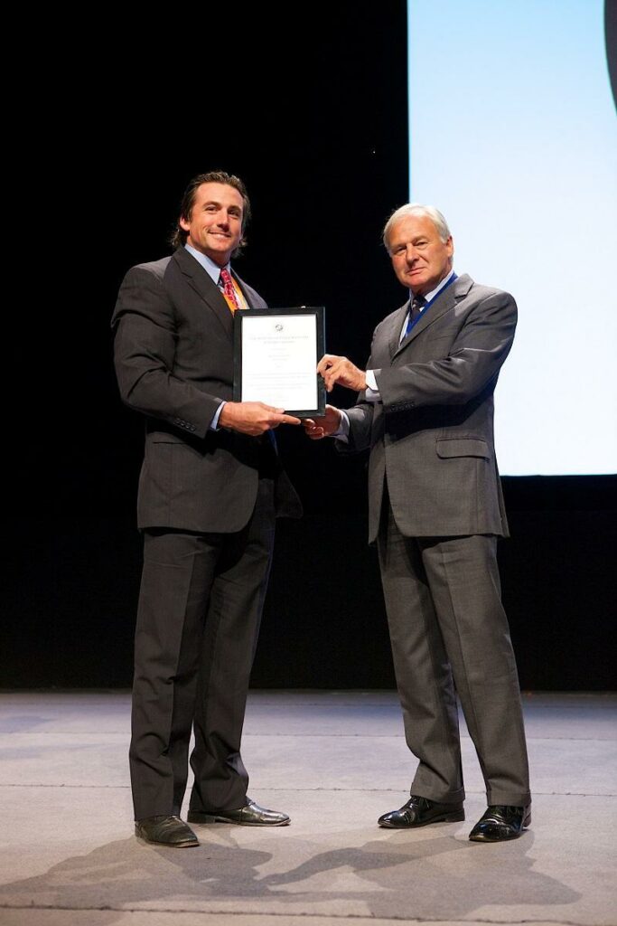
Congratulations to Dr. Weston Davis for winning the BEVA Trust Peter Rossdale EVJ Open Award at the 2014 BEVA Congress in Birmingham. The award was for a paper Dr. Davis wrote entitled:
Palm Beach Equine Clinic welcomes new veterinarian Radiologist, Dr. Sarah Puchalski, and Surgeon, Dr. Weston Davis. Dr. Puchalski is a Diplomate of the American College of Veterinary Radiology. Her specialty includes the interpretation of radiographs in addition to other diagnostic imaging techniques.
New Team Members
With Dr. Puchalski’s addition, Palm Beach Equine Clinic is pleased to be one of few practices in the country with a full-time board certified radiologist.
Dr. Davis is a Board Certified surgeon specializing in equine sports medicine. Dr. Davis joins Dr. Bob Brusie and Dr. Jorge Gomez at Palm Beach Equine Clinic providing exceptional surgical expertise in orthopedic and soft tissue related problems including emergency colic surgery.
Expanded Facilities
In addition to this exciting news comes the announcement of a new expansion to their veterinary hospital. PBEC is located less than one mile from the Winter Equestrian Festival and across the street from the Global Dressage Festival. The expansion includes a new barn containing 11 intensive care stalls capable of providing advanced medical treatment and post-operative care. The added stalls and office space makes Palm Beach Equine Clinic the most progressive, well-equipped facility in the region for equine veterinary care.
Palm Beach Equine Clinic (PBEC) is the one of the select few equine hospitals in the country with three board-certified surgeons. With 24 veterinarians on staff, they are the most complete hospital in the southeast United States, ready to handle any emergency as well as elective surgery and preventative care.
Exceptional Equine Medical Care
Palm Beach Equine Clinic has locations in Wellington, Florida, Long Island, New York and satellite veterinarians throughout the United States. PBEC provides exceptional equine medical care with an unwavering commitment to the horse for more than 30 years. Working with patients that range from the reliable amateur horse to Olympic athletes, PBEC’s mission from day one has been to provide innovative veterinary services that promote health and happiness in horses while extending their performance careers. With this mission in mind, Palm Beach Equine Clinic remains the leading service provider in equine veterinary medicine.
From December 1st, 2013 through April 1st, 2014, PBEC is pleased to offer tours by appointment. After scheduling an appointment, guests can look forward to a tour of the facility and time to talk with veterinarians. Tours will include the new hospital, Nuclear Scintigraphy room, and Surgery Center. Visitors will have an opportunity to ask questions and meet all the new doctors on staff. PBEC is located at 13125 Southfields Road in Wellington, Florida.
Contact PBEC for more information
Palm Beach Equine Clinic is respected throughout the industry. Well known for an unwavering commitment to the horse and their owners, PBEC offers innovative, advanced medical care. Known as leaders in new diagnostics and therapies, PBEC veterinarians have published numerous articles in equine magazines and journals.