Breeding the Modern Way
Palm Beach Equine Clinic’s Own Dr. Katie Atwood Discusses a 21st Century Take on Equine Reproduction
The process of breeding sport horses is ever-changing. Whether in an effort to produce the healthiest, most talented foals, to prolong the competition career of a mare, or make the most of a stallion’s longevity, reproductive science in horses has come a long way from the days of the traditional breeding shed.
Dr. Katie Atwood joined Palm Beach Equine Clinic, based in Wellington, FL, in June and brought her passion for reproductive work with her to the winter equestrian capital of the world.
“I like the creating of life,” said Dr. Atwood, who is a Florida native and University of Florida graduate and currently pursuing steps to become a board-certified reproductive specialist. “Equine medicine is intriguing, but you’re dealing with sick, unhealthy animals. With reproduction, I am working with healthy animals and making their babies, which I love!”
Embryo Transfer
The most popular wave of advancement that has hit the horse sport industry over the past several years is the process of embryo transfer.
How it works:
- A donor mare and stallion, who hold the genetics of the future foal, are bred.
- At seven or eight days of pregnancy, the embryo is flushed out.
- A catheter is placed through the vagina and cervix, and an inflatable cuff on the catheter provides a fluid-tight seal.
- A lavage fluid with surfactin (added to reduce the “stickiness” of the embryo and allow it to be extracted easily) passes down through a tubing system into the uterine lumen. As the fluid swirls throughout the lumen and drains back out through gravity, it collects the embryo, which is swept back out. The fluid and embryo pass out through the tubing system into and through an embryonic filter.
- When the embryo is identified under microscope, it is removed into a more enriched medium until the time of transfer.
- The embryo is shipped to a recipient farm where a young and healthy surrogate mare of decent size receives the embryo. That mare carries the foal to term, but it is genetically created from the donor mare and stallion.
While the process is fascinating, some may wonder why it’s necessary. According to Dr. Atwood, it relieves much of the concern owners have about breeding their sport horse mares.
“The gestation period for a horse is 11 months, so you’re only getting one foal per year when you breed traditionally,” she said. “This allows a mare to produce multiple foals per year, but it also allows that mare to remain in competition. This process can be done on younger mares with no interruptions to their competition and training schedules.”
Horses are now being bred at an ideal reproductive age while they are still in training, which is made even more valuable by the fact that advances in equine science has prolonged the longevity of horses. While 16 or 17 was once the age of an older horse, now it’s commonly seen as the age when horses are winning in the show ring. Thanks to embryo transfer, these horses can enjoy longer, healthy careers and still produce the talent of the future.
Dr. Atwood has seen embryo transfers become popular in dressage and polo, but she has begun to see it span all disciplines, saying, “At the start of the season, I had one farm and a few mares, but now it has quickly grown to several farms with multiple mares at each. It is really taking off because people now realize it does not remove their mares from competition.”
The process not only keeps mares competing, but it allows stallions to cross continents. Frozen fertilized embryos from working polo ponies in the U.S. are now being shipped to Argentina where they are carried by mares and then trained by some of the best polo trainers in the world. On the flip side, semen can also be frozen and shipped to the U.S.
“Stallions are collected, the semen is placed with an extender and high nutrient base so the sperm has something to use for energy, and then cooled slowly until it is frozen in liquid nitrogen,” said Dr. Atwood. “Once frozen, it is theoretically good forever. Last year, I bred a mare with 1991 semen and she was successfully pregnant!”
What’s Next at Palm Beach Equine Clinic
Palm Beach Equine Clinic underwent significant facility renovations over the last year, which included improvements to their onsite breeding shed. Now covered from the heat and inclement weather like an indoor arena, the shed boasts a hydraulic phantom mare.
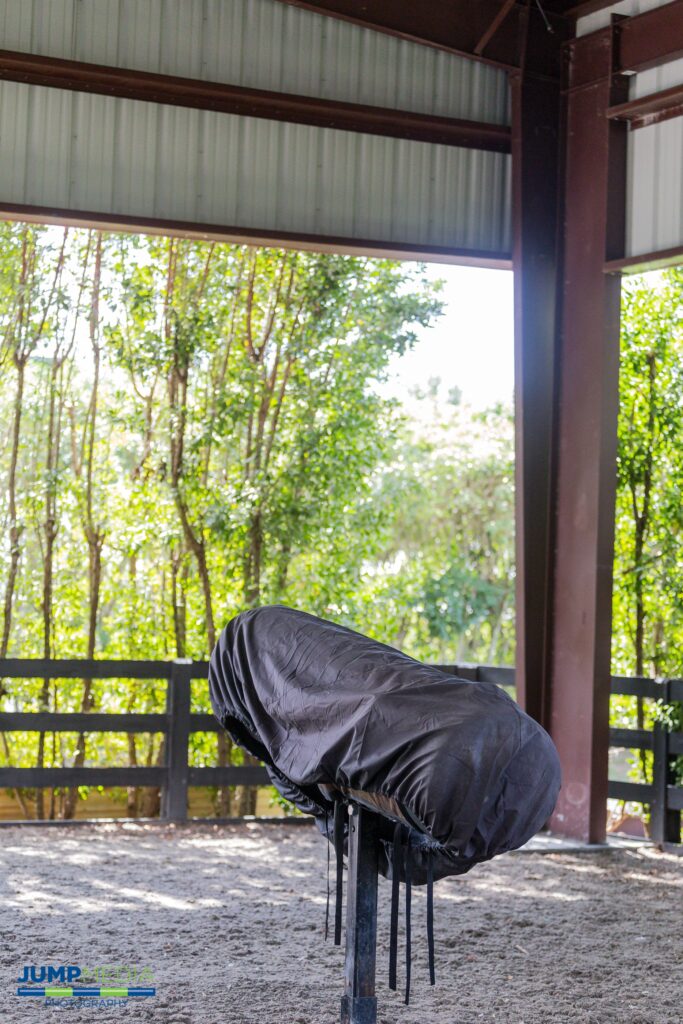
“We can raise a lower our phantom with the push of a button so it can be the appropriate for the stallion,” said Dr. Atwood. “Previously, we had to bring a tractor in to raise and lower the phantom.”
Additionally, Palm Beach Equine Clinic recently incorporated the use of a SCA® CASA (computer assisted sperm analyzer) system into their reproduction work. An excellent way to improve quality control of a stallion’s sperm, the system evaluates sperm motility (velocity and type of movement), concentration (sperm count), morphology (sperm shape), DNA fragmentation (counting of fragmented sperm), vitality (live and dead count) and acrosome reaction, which is what ultimately allows the sperm to penetrate the egg.
From onsite experience to computer technology, Palm Beach Equine Clinic offers Dr. Atwood the opportunity to be at the forefront of equine reproduction, a place she has always strived to be.
“I wanted to come into a practice that had a developed program in place, but what is even more important to me is mentoring and teaching my technicians and clients about reproduction,” she said. “It is so important to make sure these techniques are shared and promoted for the continued success of veterinarians, owners, and most of all horses.”
Speak with Dr. Atwood about breeding your horse by filling out the form below
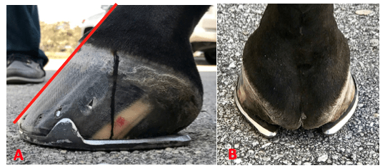
Many a seasoned horseman will admit that success in any discipline of horse sport is dependent on healthy hooves. Palm Beach Equine Clinic proudly offers the most advanced equine podiatry services to referring veterinarians and clients.

As the winter show season reaches its peak in South Florida, hoof care is paramount and the importance of good quality hoof care in the competition horse can’t be denied. The equine hoof is unique, as it is comprised of a group of biological structures that follow the laws of biomechanics. To that end, the farrier is a major asset during the show season as he or she can be proactive in maintaining the health of a horse’s foot and help to prevent lameness.
There are three very important aspects of farriery science that the farrier will use to keep any horse sound:
1. The Trim
Trimming the foot in conjunction with the size and placement of the horseshoe. Typically, a farriery session will begin with an evaluation of the conformation of each hoof from the front, side, and behind to observe the height of the heels. Next, the farrier should observe the horse in motion to see whether the horse’s foot lands heel first, flat or toe first. Regarding the trim, many farriers no longer use the term ‘balance the foot’ – which has no meaning – and have begun to use guidelines or landmarks when approaching the trim.
The guidelines used are:
- Trimming to achieve a straight hoof-pastern axis
- Using the widest part of the foot which correlates to the center of rotation
- Trimming the palmar foot (heels) to the base of the frog or to the same plane as the frog.
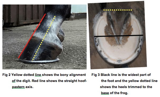
A closer look at these three guidelines, which are all interrelated, will help to show their importance. If the dorsal (front) surface of the pastern and the dorsal surface of the hoof are parallel or form a straight line, then the bones of the digit (P1, P2, P3) are in a straight line, and the force from the weight of the horse will go through the middle of the joint. Furthermore, and equally important, if the hoof-pastern axis is straight, the weight will be distributed evenly on the bottom of the foot.
2. Center of rotation (COR)
As the COR is located a few millimeters behind the widest part of each foot, it allows the farrier to apply appropriate biomechanics to each foot. The foot is trimmed in approximate proportions on either side of the widest part of the foot, which provides biomechanical efficiency.
3. The Heel

One should trim the palmar section of the foot to the base of the frog or trim such that the heels of the hoof capsule and the frog are on the same plane. Adherence to this guideline keeps the soft tissue structures (frog, digital cushion, ungula cartilages) within the hoof capsule, which are necessary to absorb concussion and dissipate the energy of impact. We must remember that heels do not grow tall, they grow forward. If we allow the heels to migrate forward, the soft tissue structures will be forced backward out of the hoof capsule. Furthermore, as the heels migrate forward, the weight is placed on the bone and lamellae, thus bypassing the soft tissue structures of the foot. Allowing the heels to migrate forward also decreases the ground surface of the foot.
These three guidelines can be applied to any foot and they serve as a basis for maintaining a healthy foot, as well as a basic starting point for applying farriery to a horse with poor foot conformation or one with a distorted hoof capsule.
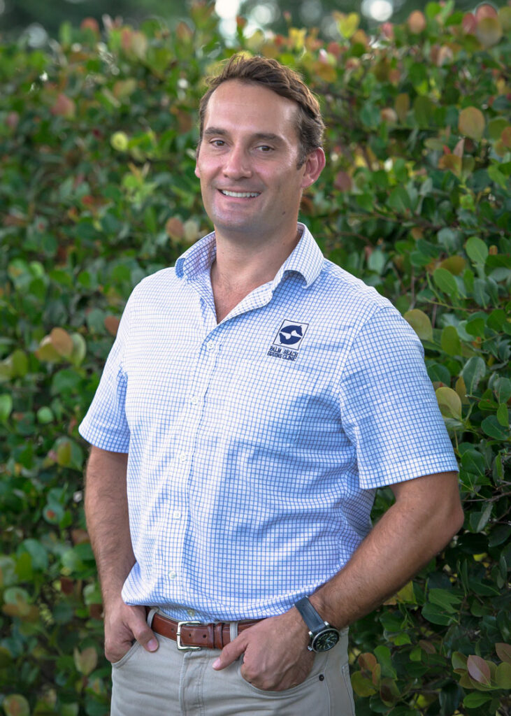
One of the repeatedly praised and recognized characteristics of Palm Beach Equine Clinic is the great pool of talented professionals from all specialties that make up the large team. In 2016, Dr. Peter Heidmann, DVM, MPH, began working with Palm Beach Equine Clinic for the winter season, and this year, the accomplished internal medicine specialist is returning to work with Palm Beach Equine Clinic for the duration of the 2018 winter season.
Dr. Heidmann graduated from Tufts University with his veterinary degree in 2000 before beginning a one-year internship in equine medicine and surgery at Arizona Equine, followed by a one-year surgical fellowship at Oregon State University, followed by a residency for internal medicine at the University of California, Davis, which he completed from 2002 to 2005. In 2005, Dr. Heidmann joined a private equine practice in Montana, and today he is the owner and hospital director of Montana Equine.
Q: What prompted you to pursue equine veterinary medicine?
I knew I wanted to be a vet for a long time, but I didn’t know that I wanted to practice more individually focused medicine until I was going through vet school. It wasn’t until my fourth year, where you’re actually starting to work with patients, that I made that realization. I started moving in that direction, and I had some faculty that saw the same for me and encouraged me to do the horse stuff. I always loved working with horses, but up until that time, I thought my background was too agricultural to do high-end sport horse work. But their encouragement helped me realize that wasn’t true. Then, I decided as a result of that realization and thought process, that I wanted to do an internship and residency to become specialized in internal medicine.
Q: What led you to Montana, and how has your role and practice evolved from when you started there in 2015 to today where you’re now the owner and hospital director of Montana Equine?
When I finished my residency at Davis in 2005, I wanted to be in the west. I wanted to be somewhere where there was nobody with my skill set – meaning internal medicine. I wanted to be in a small mountain town basically. Somewhere that I could practice the kind of medicine that I was trained to do but not in big metropolis. So, I narrowed it down to a practice in New Mexico and a practice in Montana. I went to work for a practice in Bozeman, MT, in July of 2005 and six months later my predecessor, Dr. Dave Catlin, passed away in a car crash. It was really sad. He had three kids, left a widow, and it was his dream to start an equine only specialty vet hospital, which he had done in 1999.
When he died, the conventional wisdom around Bozeman was, ‘Well this can’t be done. Dave had a dream, and it’s not a realizable dream now. There aren’t enough people here. Montana residents won’t understand specialized medicine and they won’t pay for it.’ I was faced with this proposition of leaving the practice after just arriving there, but I knew that was where I really wanted to be. I basically picked up the pieces of Dr. Catlin’s practice and formed Montana Equine in February of 2006.
Now we have a 7,500 square foot hospital with a surgery site at Bozeman, and I have a partner there, an associate, and two interns. We have two satellite locations: an ambulatory-only satellite in Helena, MT, and have nearly completed satellite clinic building in Billings, MT, which is the largest community in Montana.
Q: You began working with Palm Beach Equine Clinic during the 2016 Winter Equestrian Festival (WEF) season, but you’re out in Montana and have your own practice there. How did that relationship come about, and what does it look like today?
There are various aspects to that. We’re very quiet in Montana from basically Thanksgiving through March, and it’s not really my personality to sit around. That’s one aspect, and another is from a business perspective, it allows me to bring more vets on and keep them year-round. A third element is the desire to do more high-end medicine, and a fourth element that led me to Wellington, is my wife, Allison, who is a professional jumper rider. Two years ago, during the winter of 2015, Allison had a girlfriend who had just had a baby and asked Allison to run her business for her here at WEF. I thought that was an interesting idea and started talking to Palm Beach Equine Clinic and found that there might be a need for an internal medicine specialist in their group. So that’s when I started coming. At that point, I was in Wellington for four or five days and then home for ten days, back and forth and back and forth. Then last year, I was more of a week here and a week at home. We had our first child, so Allison and Oliver, who was brand new then, were here straight through, but I went back home to Montana as well. The need is there and the relationship is great. So, this year, I’m in Wellington full-time for the WEF.
Q: What do you enjoy most about having the opportunity to practice with Palm Beach Equine Clinic?
It’s a huge group of people, so there are a lot of personalities. When you have a lot of different personalities, you have a lot of different perspectives on treatments, and that’s interesting and fun. It means that every year when I’m here, it’s pretty dynamic. Back in Bozeman, I have a great team, but I’m the leader of the team. I’m the oldest and the most experienced. That’s good, and that’s not to say that those guys don’t definitely come with new ideas, but being here, there are so many ways of doing things that I end up picking up new tricks or new ideas even though I’ve been practicing for most of two decades. So here at Palm Beach Equine Clinic, I’m picking up new strategies and new techniques separate from new stuff coming out in the world – just different ways of doing things, and different experienced veterinarians to bounce ideas off of. That’s really refreshing and stimulating.
From a vet’s perspective, with the Wellington demographic there is sometimes less of a limitation on budget and expense, so we’re really able to set that factor and worry aside and instead focus solely on what is best for the patient. The limitation isn’t financial; the limitation is just medicine and what we are able to do, which is exciting and allows us to see great results.
Q: Have you had any favorite cases or standout moments during your time with Palm Beach Equine Clinic thus far?
I won’t name any in particular, but what I really enjoy are the challenging medical cases, the internal medicine cases that tend to be really sick and that you’re able to fix. The most common ones that we see are probably sickness after colic surgery, horses with really bad diarrhea, so colitis cases, followed by foals. Those cases generally all have a lot of things going on in a lot of different organ systems. So, there are a lot of factors to balance. Internal medicine people like me are kind of inherently nerdy, and we really get into what’s happening with the acid-base status, electrolytes, blood gases, fluid volumes, and other technical aspects of horse medicine.
Q: What do you enjoy doing outside of work?
While in South Florida, we’re basically working all the time, so not as much here except trying to get exercise and be fit. I like to ride a bike or go for a run. At home, the Gallatin Valley – which is where Bozeman is – is surrounded all around by mountains. The valley floor at Bozeman is at about 5,000 feet, and the peaks are at 8,500, so you can do a lot of really serious hiking. Our son is only 18 months old, but he loves it. I’ve had dogs in the past where their whole personality changes when you get out of town, and that’s what Oliver is like too. It’s almost like his whole face is a different face when he’s out there.
Any horse owner’s worst nightmare is realized when their mount begins to show the dreaded signs of colic. For Jody Stoudenmier, a Wellington, FL, resident and avid dressage rider, she knows the symptoms all too well.

Patient History
Stoudenmier owns an 11-year-old American-bred Dutch Warmblood mare that joined her string of horses at the end of 2016 and has competed through the Intermediate II level. Sidelined by a suspensory injury last year, Beatrix was prescribed stall rest to aid in her recovery by Dr. Robert Scott of Scott Equine Services based in Ft. Lauderdale, FL. An unfortunate but common side effect of the necessary stall rest was colic. Beatrix suffered from six bouts of colic that were resolved without surgery when Dr. Scott referred Stoudenmier and Beatrix to Palm Beach Equine Clinic.
“She is such a wonderful mare; a nice mover, very athletic, sweet, sensitive, and easy to handle in the barn,” said Stoudenmier of the mare that regularly competes at the Adequan Global Dressage Festival during the winter season. “When she was recovering from her injury we tried everything to prevent her from colicking – diet, medications, hand walking – but, nothing seemed to be working.”
It was then that Palm Beach Equine Clinic’s board-certified surgeon, Dr. Weston Davis, suggested a laparoscopic surgical approach.
“Her colic had never progressed so far that we needed to do surgery before,” explained Stoudenmier. “But, at that point, I was open to anything! After speaking with Dr. Davis, I immediately had a positive feeling about it.”
Dr. Davis’ Surgical Procedure
The procedure that Dr. Davis suggested was an endoscopic ablation of nephrosplenic space. In layman’s terms, as a result of Beatrix’s colic, her colon was essentially getting caught or entrapped over the nephrosplenic ligament, which connects the left kidney to the spleen. When the colon is entrapped in this position, its contents cannot move through it and the colon becomes distended, causing the horse considerable pain, and the inevitable colic.
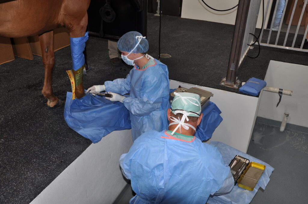
Dr. Davis’ solution was to close or perform an ablation of the nephrosplenic space to prevent further entrapment. The procedure can be conducted endoscopically where the horse does not have to be anesthetized, but undergoes a standing surgery with sedation and local anesthesia. A small incision is made in the left flank and the laparoscope is inserted through a smaller incision close by. The nephrosplenic space is then sutured closed so that the trough that forms the space between the kidney and spleen is obliterated and can no longer entrap the colon.
On October 9, 2017, Beatrix underwent a successful ablation of the nephrosplenic space at the hands of Dr. Davis.
“In the past, I have had several horses undergo surgery where they had to be anesthetized and it was very difficult to get them standing again after surgery,” said Stoudenmier. “We did not have that worry with Beatrix and the approach absolutely made a difference in her recovery.”
Post-Surgery Care and Recovery
Beatrix remained at Palm Beach Equine Clinic for a week and a half after surgery to jump-start her recovery before returning home to Stoudenmier, who has managed her post-surgery care with the help of both Dr. Davis and Dr. Scott.
“Dr. Davis was absolutely wonderful to work with,” said Stoudenmier of her experience at Palm Beach Equine Clinic. “He listened to my concerns, was patient, and kept his mind open.”
Dr. Davis paid a visit to Beatrix in mid-November to perform an ultrasound and together with Dr. Scott approved the mare to return to work. Stoudenmier has begun to introduce trot work into Beatrix’s routine and is optimistically expecting a full recovery, saying, “She looks super and everything looks good for the next two months. My goal by the end of the season is to get her back in the show ring!”
Meet PBEC Veterinarian Dr. Natalia Novoa
Dr. Natalia Novoa was born in Nashville, TN, and raised in Colombia, where she graduated from the University of La Salle. Upon returning to the United States, Novoa validated her degree in both the U.S. and Canada and received her chiropractic adjustments qualifications in Kansas and studied acupuncture in Florida. Novoa joined the Palm Beach Equine Clinic (PBEC) team for a short period in 2000, before spending several years following the hunter/jumper horse show circuit in Canada, Colorado, and Ocala, FL. She then re-joined the PBEC staff in 2011, and, with her veterinary license in Florida, Kentucky, North Carolina, and Canada, Novoa has been a valued member of their team ever since.
Q: How did you decide that you wanted to pursue veterinary medicine?
I have enjoyed having animals in my life since I was very young, but what was key to me deciding to pursue veterinary medicine was having a farm where I was able to gain skills and real-world experience that would prove handy down the road. I was able to practice with real medical cases and acquire knowledge in operational management, and it was an introduction to the lifestyle because it requires patience, hard work, commitment, determination, and accountability. It was a valuable opportunity to work hands-on in the environment with the veterinarian of my farm, absorbing, assimilating, and getting familiarized with the vet skills that were the building blocks for my future. He made me realize that veterinary medicine would fit me perfectly. That helped me set the foundation for my career, and since then, I didn’t have to look any further!
Q: What led you to PBEC?
I think of it like this: if I want to become the best at what I do then I have to work with the best, and I picked PBEC. They say, “Leadership is not about a title or designation. It’s about impact, influences, and inspiration.” Palm Beach Equine assumes the leadership role, chases the latest trends, and evolves continuously. In addition, we have a great team of skilled vets and staff who try to expand their vision and abilities to achieve success.
I have to say I have had seven great horse show seasons with Palm Beach Equine, and I’m looking forward to an eighth one. When you are doing what you love and you are working with great horses, time flies.
Q: What is your primary area of focus?

I focus on sports medicine and lameness, combining conventional medicine with alternative medicine; this includes chiropractic adjustments, acupuncture, and herbal therapy. To me those are great alternative tools for diagnosis and treatment. I have to say, they help you to look at a horse or a dog in a different way, and you have more tools and options. Regardless of the case – lameness, pain management, internal medicine, etc. – I have different choices to achieve results in those situations.
I perform chiropractic adjustments, acupuncture therapy, and laser treatments to small animals as well. Most horse people have dogs, so I’m able to take care of their companions too.
Q: What is a typical day like for you at PBEC?
It’s a busy agenda every day with continuous juggling. I’m usually dealing with lameness (involving diagnosis and treatment), doing pre-purchase exams, chiropractic adjustments, acupuncture therapies on horses and dogs, performing laser treatments, and at the end of the day, doing paperwork. Depending on the day, I could be doing FEI treatments, vet shift at the horse show, donating vet work at Vinceremos Therapeutic Riding Center, or dealing with an emergency. We’re a very mobile practice able to go from farm to farm and horse show to horse show (Kentucky; Tryon, NC; Ocala, FL), so I’m moving around a lot.
Q: What is your favorite part of the job?
There are so many aspects that I enjoy: working outdoors, closely with animals on a daily basis, and diagnosing and dealing with different issues to overcome problems; the privilege of working as a team with the clients and trainers and creating a great, close relationship with them; the connection with the animals and how they show you how grateful they are, and being able to see the improved outcome. There’s great satisfaction in successfully applying my knowledge and skills to a make a difference in someone’s life.

Q: Have you had any standout, favorite moments since beginning to work at PBEC?
When I had double success treating a ‘mystery’ case of a horse in which the owner was losing hope because other treatments were not showing good outcomes, and also treating her dog that was crying and unable to get in the car. The animals and owners were both happy and thankful. “For it is in giving that we receive” – having a positive impact on someone is correlated with high levels of overall job satisfaction.
Q: What do you enjoy doing outside of work?
I enjoy traveling with my daughter, Lola, and visiting my parents and siblings that live in different countries. I also love dancing to Latin music, running, playing tennis and squash. I’m a pretty competitive squash player, so I welcome any challengers!
Fear of colic is in the back of many horse owners’ minds, but with the expert care of Palm Beach Equine Clinic, owners can rest easy knowing that they have some of the world’s best surgeons and veterinarians at their disposal in the event of an emergency.
Colic 101
Characterized by abdominal pain or problems with the gastrointestinal tract, colic is something that often arises unexpectedly and from many different origins. Spoiled feed, abrupt changes in feed, parasite infestation, sand ingestion, lack of water consumption, and even excess stress or changes in the weather are among the numerous causes generally related to colic.
Colic Symptoms
Whatever the cause may be, the most important step any owner can take is to recognize the symptoms as early as possible and immediately call their veterinarian. Pawing, rolling, looking at abdomen, sweating, loss of interest in food and water, and absence of gut sounds in any of the four abdominal quadrants are some of the telltale signs of colic development. Unfortunately, colic can be fatal, but the proper knowledge and care may save your horse’s life. The sooner your veterinarian gets involved in treatment, the better your horse’s chance of survival.
Emergency Colic Care
In the event of an emergency, the veterinarians and surgeons of Palm Beach Equine Clinic are available 24/7 to offer the very best care for your equine partner. Palm Beach Equine Clinic is renowned for its referral full-service surgical center and intensive care hospital located in the heart of Wellington, Florida. Board-Certified surgeons, primary care veterinarians, and skilled hospital technicians are available to treat, monitor, and care for critical cases. With world-class veterinarians and a full staff of highly trained technicians, both clients and patients of Palm Beach Equine Clinic are in the best hands possible.
Surgical Capabilities
Palm Beach Equine Clinic offers the latest in technology as the surgical techniques are less invasive and result in faster recovery times for your horse. The surgical team leader, Dr. Robert Brusie, is a nationally renowned, Board-Certified surgeon. Dr. Brusie’s surgical specialties include orthopedic, arthroscopic, and emergency cases. Dr. Brusie has been the head surgeon with PBEC for the past 20 years.
“In the last ten years, colic surgery has come a long, remarkable way,” Dr. Brusie stated. “With our clients, if the horse needs to go to surgery, we get an approximately 95% success rate. We attribute that to the client’s excellent care of their horses, as well as their knowledge to contact us immediately. That being said, colic surgery is always the last resort. We try to help all horses improve medically first.”
Palm Beach Equine’s surgical suite and staff is prepared to handle all types of emergencies, day and night. The large team of 40 veterinarians includes three Board-Certified Surgeons who rotate on-call duties so every day is covered. This aids Palm Beach Equine Clinic veterinarians and all of Southeast Florida with the ability to treat their emergencies requiring surgical assistance as quickly as possible. The state-of-the-art intensive care hospital is equipped with top-of-the-line medical equipment, including digital video cameras for the clinicians to easily monitor their patients from any location, at any time.
Meet Surgical Resident Dr. Michael Myhre
Dr. Michael Myhre was born to be a veterinarian. In 1978 his father, Dr. Grant Myhre, developed a referral practice, Myhre Equine Clinic in Rochester, NH. After working alongside his father since middle school, Dr. Myhre, who hails from Milton, NH, believes he was always destined to be a veterinarian. Dr. Myhre graduated from Cornell University College of Veterinary Medicine based in Ithaca, NY, in 2016, and he joined Palm Beach Equine Clinic thereafter as a surgical resident to work under the direction of board-certifed surgeons Dr. Robert Brusie, Dr. Jorge Gomez, and Dr. Weston Davis.
What is your background with horses?
I grew up in my father’s practice. He would bring me along to see outpatients and cut colics at 2 a.m. When I was in high school and college, I would work there during the summers as a technician. I kept learning from him and when it was time to decide what I would do, I applied to vet school.
We had some lesson horses at home and taught some therapeutic riding, so I rode on the trails occasionally, but I knew I was always supposed to be a veterinarian.
Where did you complete your undergraduate degree?
I attended Ithaca College in New York and studied computer science. It is a pretty unusual undergraduate degree for a veterinarian, but I did not want to go the traditional route of getting a biology degree. Computer technology is now involved in a lot of veterinary medicine – so much of what we do is going through computers, so it is an asset to have that degree.

I still took all the biology and chemistry classes at the same time, and I finished in three years. At that point, I applied to Cornell University and was accepted.
What led you to Palm Beach Equine Clinic?
I came here because it is the best residency program in the country. I have a big caseload and get to work on the best horses in the world. I started on July 1 and what I like the most is the diversity in cases. We have seen hunters, jumpers, dressage horses, and racehorses. I have done everything from condylar fracture repairs to MRIs, nuclear scintigraphy, x-rays, and even colic surgery on a miniature horse. Palm Beach Equine Clinic stays at the forefront of technology with a new standing surgery pit, standing MRI machine, and paperless medical records.
What goals do you have for your veterinary career?
After my three-year residency at Palm Beach Equine Clinic, I plan to move back to New Hampshire and work at my father’s practice.
What can we find you doing when you’re not working?
I am pretty much always working, but my girlfriend is a neurology resident in Manhattan, so I try to visit her as much as I can, or I take advantage of living in Florida and go swimming.
Name one thing most people wouldn’t know about you?
I rowed for the Ithaca College crew team and while I was in vet school, I was an assistant coach for the Cornell University team.
Success Story: Freeman
In January 2016, the Pine Hollow team noticed something seemed off just before driving out of the Winter Equestrian Festival (WEF) with their horses. Stopping to check the horses before continuing off the showgrounds, Pine Hollow discovered Freeman, a promising and successful Dutch Warmblood, had swung his hind leg over the back of the trailer. Freeman’s stifle had ended up squarely on one of the hooks used to secure the back door, lodging the hook into his stifle and into the femoropatellar joint.
Emergency Veterinary Care
Recognizing the extreme peril facing Freeman, Pine Hollow immediately called for help from Palm Beach Equine Clinic, the Official Veterinarians of WEF.
“It took tremendous effort, creative thinking, and exceptional teamwork to free Freeman from the hook impaling his leg,” said David Blake, Pine Hollow’s internationally acclaimed rider and trainer. “Palm Beach Equine Clinic sent several of their top vets to help us rescue Freeman. The team of vets is truly great.”
Thanks in very large part to the help and determination of the vets, Pine Hollow and Palm Beach Equine Clinic were able to free Freeman from the trailer door.
At the Equine Hospital
From there, Freeman was transported to the nearby Equine Hospital, where he spent a few days recovering before it was agreed to pursue arthroscopic surgery on his femoropatellar joint.
“To be honest, it wasn’t looking good at all for the first day or so Freeman was there,” said Blake. “The joint was so severely damaged we didn’t know if it could be fixed. Our only chance of fixing the joint was surgery, so we agreed we would try everything possible.”

Dr. Weston Davis performed the surgery, after which Freeman remained in Palm Beach Equine Clinic’s care while he regained use of the leg.
“The team did a fantastic job there and kept Freeman until he was ready to begin long-term rehab with James Keogh,” said Blake.
When Freeman was finally ready to return home to Pine Hollow, Blake hoped at best Freeman would eventually be able to do light work and perform at a low level.
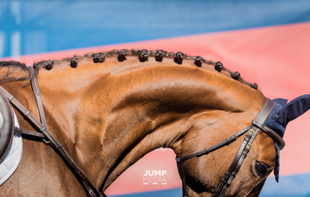
In the past, when a horse’s gait has felt off or lacking in its usual impulsion, it was often assumed to be an issue of lameness. Now however, thanks to the improved diagnostics readily available at the Palm Beach Equine Clinic veterinarians are able to more accurately pinpoint the problem area. Perhaps surprisingly, it’s not always in the legs or hooves. With increasing frequency, the horse’s neck is being diagnosed as the root of the issue.
The Anatomy of the Equine Neck and What Can Go Wrong
In order to understand the problems that can arise in association with the horse’s neck, it’s important to first understand the anatomy.
The neck is composed of seven cervical vertebrae running from the head to the thorax, named C1 through C7, and each articulating with each other. The primary purposes of the neck are to move the head and to protect and transport the spinal cord and nerves, which run through the middle of the vertebrae.
Such a major role as the protection of the nerves and spinal cord can also come with some major risks and complications, with clinical signs of these problems generally presenting themselves either neurologically, as neck pain, or as lameness in the front legs. These more specific symptoms may include:
- Ataxia or clumsiness – Ataxia is defined as the “lack of control of bodily movements”. In the case of an ataxic horse, you may begin to notice staggering, sudden loss of balance, or even an inability to remain upright. Ataxia is generally an indicator of a neurological condition or damage to the spinal cord itself, caused by either developmental issues, trauma, or an infectious disease such as equine protozoal myeloencephalitis (EPM). Such neurological cases can often be the most debilitating.
- Lameness – You can think of the spinal cord and the nerves in the neck like an interstate, with the spinal cord itself acting as the major highway. As you are “driving” along the interstate, every so often there are little exits, which is where the other nerves come out. Should there be any impingement on the interstate or spinal cord itself, you’re likely looking at more severe complications – much like an accident on the highway. Should there be impingement on the nerves coming off of the spinal cord, it will more likely present itself like an accident a little way off an exit – not affecting the interstate itself, but possibly causing problems that spread elsewhere. That is where we see lameness issues arise.
This can be more difficult to pin down, but can often be due to pressure on the smaller nerves that pass through the openings in the vertebrae and supply the front legs. Arthritis of the articular facet joints of the vertebrae is another common reason for lameness, as anytime these joints become arthritic or inflamed, it can easily translate to the forelimbs.
- Neck Pain – This will often go hand-in-hand with lameness, as factors such as arthritis of the articular facet joints can lead to both symptoms. Other possible reasons for neck pain include trauma or inflammatory diseases.
Diagnosing the Problem
Neck problems, particularly those related to lameness, are generally diagnosed through a process of exclusion, first performing nerve blocks to or ruling out lower regions of the horse’s body. Palpation of the neck, testing of the neck’s movement, and full neurological exams may also be performed in addition to a full lameness exam, depending on the horse’s symptoms.
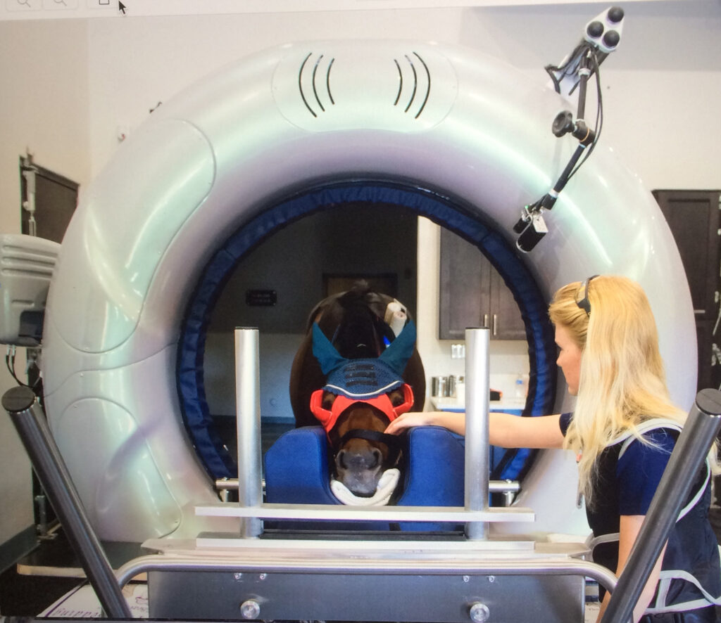
Once other regions of the horse are ruled out as the location of the problem, veterinarians are now able to use diagnostic images such as radiographs, nuclear scintigraphy and standing CT scans to specifically locate problems in the neck like never before.
In years past, those diagnostic resources were left for last-ditch cases when veterinarians really could not pinpoint any other problems. Today, with the advent of more modern technology, better radiographs, better ultrasound machines, and the more advanced imaging of nuclear scintigraphy and CT scans, veterinarians are able to readily utilize advanced diagnostics to save time and money, and to find the root of the problem more quickly and accurately.
The neck is one of the areas that has most clearly benefited from the progression in advanced imaging. While neck problems have likely been prevalent for some time, veterinarians are now finding those diagnoses more common, as they are able to more accurately locate the issue – particularly at clinics like Palm Beach Equine Clinic. Often these issues will go undiscovered or undiagnosed in the field, but they can be identified at Palm Beach Equine Clinic thanks to the readily available imaging tools.
As an example, if arthritic factors are suspected, nuclear scintigraphy can be used to look for areas of increased bone turnover. On the resulting bone scan, veterinarians are looking for areas where there is an increased calcium uptake because the bone is actively remodeling. These areas will appear darker on the scan and are generally a good indicator of a boney injury or arthritis, with the darker “hot spots” often appearing above the articular facet joints.
Also new and groundbreaking for the diagnosis of equine neck problems is the use of a Computerized Tomography or CT scan, with the ability to scan a standing horse with light sedation on the near horizon which will be available at the clinic this upcoming winter season.
Treatment
Once a solid diagnosis is arrived upon, the proper treatment protocols can be prescribed. Depending on the root of the problem, possible treatments may include shockwave therapy, regenerative therapies such as interleukin-1 receptor antagonist protein (IRAP) therapy or platelet-rich plasma (PRP) therapy, or one of the most common treatments, injections of the facet joints.
In the case of facet joint injections, veterinarians at Palm Beach Equine Clinic are able to medicate under ultrasound guidance, guiding a needle into the joints and delivering corticosteroids or similar medication. Surgery is also an option as a final approach to severe complications.
In milder cases, treatments may also just call for increased time off, chiropractic treatments, or the administration of non-steroidal anti-inflammatory (NSAID) medications.
If you suspect any issues with your horse’s neck, contact Palm Beach Equine Clinic any time by calling (561) 793-1599 to schedule an appointment.
This month, Palm Beach Equine Clinic welcomed a new face to their team. Dr. Katie Atwood, hails from Jacksonville, FL, and attended veterinary school at the University of Florida, making her return to south Florida from Lexington, KY, a special homecoming.
What brought you to Palm Beach Equine Clinic?
I grew up in Florida, so I wanted to be closer to family and the ocean! But, I was also looking for an opportunity to grow and become a better veterinarian. This is a difficult industry to get into, but it is especially difficult to find the right practices. This is a chance for me to work with some of the best doctors in the country.
What would you say is your specialty at Palm Beach Equine Clinic?
In addition to general, preventative and sport medicine, I will be focusing on Palm Beach Equine Clinic’s reproductive work. I did an internship and a fellowship in reproduction and realized that it is what I am most interested in. I will be working on breeding mares, performing frozen and fresh semen inseminations, as well as breeding management and embryo flushes for transfers to recipient mares.
What inspired you to be a veterinarian?
When I was a little kid we had a trail behind our house that was really popular and I would sit on the back wall and watch everybody ride by on their horses. We do not have any other veterinarians in my family, but when I was five years old I realized that I wanted to work with animals. During my undergraduate studies in Animal Science at Berry College in Rome, GA, a professor named Dr. Martin Goldberg really pushed me to pursue veterinary school.
I wake up every morning so excited to go to work and if I don’t come home exhausted and filthy then I have done something wrong. It is an “every minute of every day” commitment, but very rewarding.
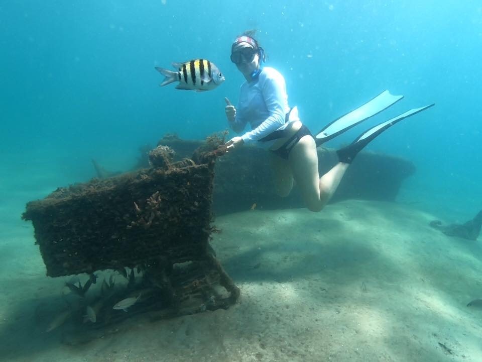
When you aren’t working, where can we find you?
I like to spend as much time as possible in the water. I can usually be found swimming, diving or paddleboarding at the beach and spending time by the pool
What advice would you give to someone considering pursuing veterinary school?
Do it! It will be the most difficult time of your life, but if you have a passion for it, it will be so rewarding. Dedication is so important; take advantage of every wet lab you can, go to any conference that is available, and take advantage of opportunities to meet new people and gain mentors. Who wouldn’t want to do what they love for a living?
Name one thing most people wouldn’t know about you?
I am a pretty open book at this point. But, when I retire, my fiancé Mackenzie and I want to sail around the world!