Category: Medical
As sport horses become faster and stronger, veterinary medicine is often challenged to break barriers to provide the best in diagnostic and maintenance care. Palm Beach Equine Clinic is consistently on the forefront of those advances and employs a team of veterinarians equipped with the latest developments in regenerative medicine.
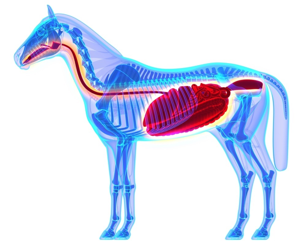
Two resources that have become increasingly popular to treat equine injuries are Platelet Rich Plasma (PRP) and Interleukin-1 Receptor Antagonist Protein (IRAP) to encourage regeneration of injured or degenerative tissue. Managing joint diseases and injuries using these methods is ground-breaking, but logical at their core. They essentially use naturally-occurring proteins, cells, and other natural bodily processes. Regenerative therapies put the horse’s own biological mechanisms to work stimulating healing without the use of steroids or other drugs.
What is PRP?
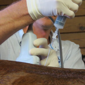
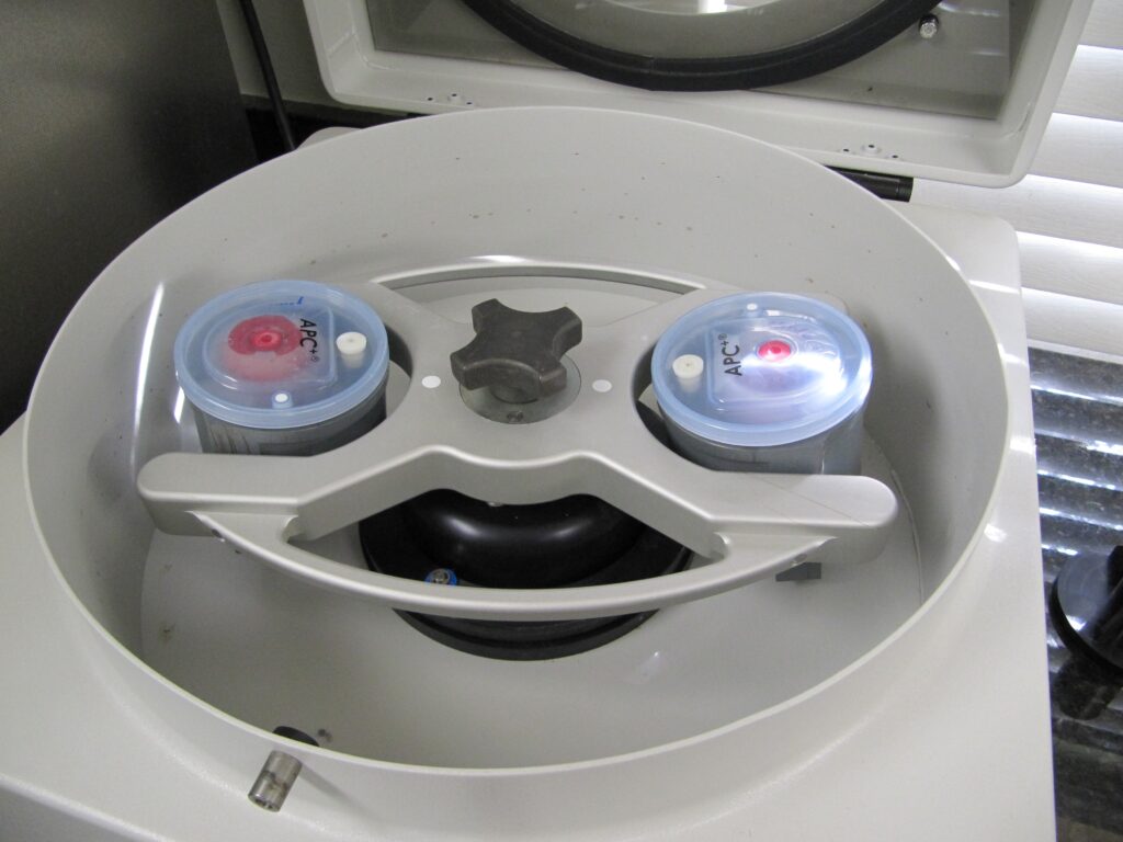
Platelets are among the very first cells to accumulate at an injured site, making them very important when simulating the repair process. Platelets contain granules filled with growth factors (the elements that aid in healing) and stimulate specified tissue to heal at an increased rate. To treat a horse with PRP, the veterinarians at PBEC are able to take a sample of the horse’s blood and concentrate the platelets in a high-speed centrifuge onsite. The harvest and processing procedure takes approximately 30 minutes before the concentrated platelet rich sample is injected back into the horse at the specific area of injury using sterile techniques and guided by ultrasound.
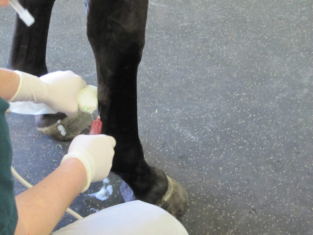
PBEC’s Board-Certified Staff Surgeon, Dr. Weston Davis, explained PRP use in more detail: “We harvest a large quantity of blood, anywhere from 60 to180 milliliters, and we process that to concentrate the segment that is very rich in platelets. We get a high concentration of platelets – we are hoping for five to eight times the concentration that you would get from normal blood. Then we take that platelet-rich extract and inject it back into an injured area to encourage a more robust healing response. Whenever you have an injury, platelets are one of the first cells that get there. They will aggregate, clump, and de-granulate. They release these granules, which are very rich in growth factors, and signal the body to start the healing process.”
What is IRAP?
IRAP is used to treat equine athletes that are susceptible to musculoskeletal injuries and osteoarthritis or degenerative joint disease. Joint trauma results in the release of inflammatory mediators such as Interleukin-1 (IL-1). IRAP uses a horse’s own anti-inflammatory protein found within the blood to counteract the destructive effects of IL-1 to slow the process of osteoarthritis. The process works by binding to the IL-1 receptors in the joint and blocking the continuation of damage and inflammation.
“We often see joint damage in sport horses because of the nature of their work, but we try to avoid overuse of steroids in joints because steroids can have long term effects on cartilage,” said PBEC veterinarian Dr. Samantha Miles. “This is a way we can manage joint disease and stop inflammation without having to consistently use steroids every time. Some of our clients will maintain their horses on IRAP alone for joint injections.”
The goal to better serve sports horses that continue to improve athletically is the driving force behind the development of even more developed and precise techniques used in regenerative medicine. And, at PBEC, the work to break new ground is never finished.
“I believe we are learning more about these technologies with more advanced science behind what they do and how they do it,” said Miles. “These treatments are natural, drug-free, and competition safe, and necessity drives the need for regenerative therapies in the sport horse world.”
Breeding the Modern Way
Palm Beach Equine Clinic’s Own Dr. Katie Atwood Discusses a 21st Century Take on Equine Reproduction
The process of breeding sport horses is ever-changing. Whether in an effort to produce the healthiest, most talented foals, to prolong the competition career of a mare, or make the most of a stallion’s longevity, reproductive science in horses has come a long way from the days of the traditional breeding shed.
Dr. Katie Atwood joined Palm Beach Equine Clinic, based in Wellington, FL, in June and brought her passion for reproductive work with her to the winter equestrian capital of the world.
“I like the creating of life,” said Dr. Atwood, who is a Florida native and University of Florida graduate and currently pursuing steps to become a board-certified reproductive specialist. “Equine medicine is intriguing, but you’re dealing with sick, unhealthy animals. With reproduction, I am working with healthy animals and making their babies, which I love!”
Embryo Transfer
The most popular wave of advancement that has hit the horse sport industry over the past several years is the process of embryo transfer.
How it works:
- A donor mare and stallion, who hold the genetics of the future foal, are bred.
- At seven or eight days of pregnancy, the embryo is flushed out.
- A catheter is placed through the vagina and cervix, and an inflatable cuff on the catheter provides a fluid-tight seal.
- A lavage fluid with surfactin (added to reduce the “stickiness” of the embryo and allow it to be extracted easily) passes down through a tubing system into the uterine lumen. As the fluid swirls throughout the lumen and drains back out through gravity, it collects the embryo, which is swept back out. The fluid and embryo pass out through the tubing system into and through an embryonic filter.
- When the embryo is identified under microscope, it is removed into a more enriched medium until the time of transfer.
- The embryo is shipped to a recipient farm where a young and healthy surrogate mare of decent size receives the embryo. That mare carries the foal to term, but it is genetically created from the donor mare and stallion.
While the process is fascinating, some may wonder why it’s necessary. According to Dr. Atwood, it relieves much of the concern owners have about breeding their sport horse mares.
“The gestation period for a horse is 11 months, so you’re only getting one foal per year when you breed traditionally,” she said. “This allows a mare to produce multiple foals per year, but it also allows that mare to remain in competition. This process can be done on younger mares with no interruptions to their competition and training schedules.”
Horses are now being bred at an ideal reproductive age while they are still in training, which is made even more valuable by the fact that advances in equine science has prolonged the longevity of horses. While 16 or 17 was once the age of an older horse, now it’s commonly seen as the age when horses are winning in the show ring. Thanks to embryo transfer, these horses can enjoy longer, healthy careers and still produce the talent of the future.
Dr. Atwood has seen embryo transfers become popular in dressage and polo, but she has begun to see it span all disciplines, saying, “At the start of the season, I had one farm and a few mares, but now it has quickly grown to several farms with multiple mares at each. It is really taking off because people now realize it does not remove their mares from competition.”
The process not only keeps mares competing, but it allows stallions to cross continents. Frozen fertilized embryos from working polo ponies in the U.S. are now being shipped to Argentina where they are carried by mares and then trained by some of the best polo trainers in the world. On the flip side, semen can also be frozen and shipped to the U.S.
“Stallions are collected, the semen is placed with an extender and high nutrient base so the sperm has something to use for energy, and then cooled slowly until it is frozen in liquid nitrogen,” said Dr. Atwood. “Once frozen, it is theoretically good forever. Last year, I bred a mare with 1991 semen and she was successfully pregnant!”
What’s Next at Palm Beach Equine Clinic
Palm Beach Equine Clinic underwent significant facility renovations over the last year, which included improvements to their onsite breeding shed. Now covered from the heat and inclement weather like an indoor arena, the shed boasts a hydraulic phantom mare.
“We can raise a lower our phantom with the push of a button so it can be the appropriate for the stallion,” said Dr. Atwood. “Previously, we had to bring a tractor in to raise and lower the phantom.”
Additionally, Palm Beach Equine Clinic recently incorporated the use of a SCA® CASA (computer assisted sperm analyzer) system into their reproduction work. An excellent way to improve quality control of a stallion’s sperm, the system evaluates sperm motility (velocity and type of movement), concentration (sperm count), morphology (sperm shape), DNA fragmentation (counting of fragmented sperm), vitality (live and dead count) and acrosome reaction, which is what ultimately allows the sperm to penetrate the egg.
From onsite experience to computer technology, Palm Beach Equine Clinic offers Dr. Atwood the opportunity to be at the forefront of equine reproduction, a place she has always strived to be.
“I wanted to come into a practice that had a developed program in place, but what is even more important to me is mentoring and teaching my technicians and clients about reproduction,” she said. “It is so important to make sure these techniques are shared and promoted for the continued success of veterinarians, owners, and most of all horses.”
Speak with Dr. Atwood about breeding your horse by filling out the form below
Many a seasoned horseman will admit that success in any discipline of horse sport is dependent on healthy hooves. Palm Beach Equine Clinic proudly offers the most advanced equine podiatry services to referring veterinarians and clients.

As the winter show season reaches its peak in South Florida, hoof care is paramount and the importance of good quality hoof care in the competition horse can’t be denied. The equine hoof is unique, as it is comprised of a group of biological structures that follow the laws of biomechanics. To that end, the farrier is a major asset during the show season as he or she can be proactive in maintaining the health of a horse’s foot and help to prevent lameness.
There are three very important aspects of farriery science that the farrier will use to keep any horse sound:
1. The Trim
Trimming the foot in conjunction with the size and placement of the horseshoe. Typically, a farriery session will begin with an evaluation of the conformation of each hoof from the front, side, and behind to observe the height of the heels. Next, the farrier should observe the horse in motion to see whether the horse’s foot lands heel first, flat or toe first. Regarding the trim, many farriers no longer use the term ‘balance the foot’ – which has no meaning – and have begun to use guidelines or landmarks when approaching the trim.
The guidelines used are:
- Trimming to achieve a straight hoof-pastern axis
- Using the widest part of the foot which correlates to the center of rotation
- Trimming the palmar foot (heels) to the base of the frog or to the same plane as the frog.
A closer look at these three guidelines, which are all interrelated, will help to show their importance. If the dorsal (front) surface of the pastern and the dorsal surface of the hoof are parallel or form a straight line, then the bones of the digit (P1, P2, P3) are in a straight line, and the force from the weight of the horse will go through the middle of the joint. Furthermore, and equally important, if the hoof-pastern axis is straight, the weight will be distributed evenly on the bottom of the foot.
2. Center of rotation (COR)
As the COR is located a few millimeters behind the widest part of each foot, it allows the farrier to apply appropriate biomechanics to each foot. The foot is trimmed in approximate proportions on either side of the widest part of the foot, which provides biomechanical efficiency.
3. The Heel

One should trim the palmar section of the foot to the base of the frog or trim such that the heels of the hoof capsule and the frog are on the same plane. Adherence to this guideline keeps the soft tissue structures (frog, digital cushion, ungula cartilages) within the hoof capsule, which are necessary to absorb concussion and dissipate the energy of impact. We must remember that heels do not grow tall, they grow forward. If we allow the heels to migrate forward, the soft tissue structures will be forced backward out of the hoof capsule. Furthermore, as the heels migrate forward, the weight is placed on the bone and lamellae, thus bypassing the soft tissue structures of the foot. Allowing the heels to migrate forward also decreases the ground surface of the foot.
These three guidelines can be applied to any foot and they serve as a basis for maintaining a healthy foot, as well as a basic starting point for applying farriery to a horse with poor foot conformation or one with a distorted hoof capsule.
Fear of colic is in the back of many horse owners’ minds, but with the expert care of Palm Beach Equine Clinic, owners can rest easy knowing that they have some of the world’s best surgeons and veterinarians at their disposal in the event of an emergency.
Colic 101
Characterized by abdominal pain or problems with the gastrointestinal tract, colic is something that often arises unexpectedly and from many different origins. Spoiled feed, abrupt changes in feed, parasite infestation, sand ingestion, lack of water consumption, and even excess stress or changes in the weather are among the numerous causes generally related to colic.
Colic Symptoms
Whatever the cause may be, the most important step any owner can take is to recognize the symptoms as early as possible and immediately call their veterinarian. Pawing, rolling, looking at abdomen, sweating, loss of interest in food and water, and absence of gut sounds in any of the four abdominal quadrants are some of the telltale signs of colic development. Unfortunately, colic can be fatal, but the proper knowledge and care may save your horse’s life. The sooner your veterinarian gets involved in treatment, the better your horse’s chance of survival.
Emergency Colic Care
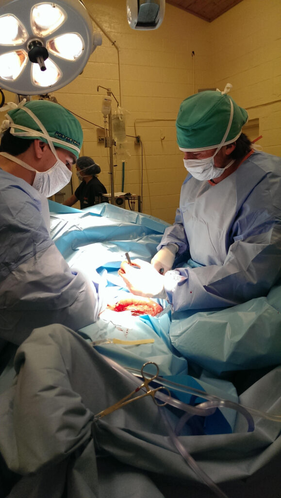
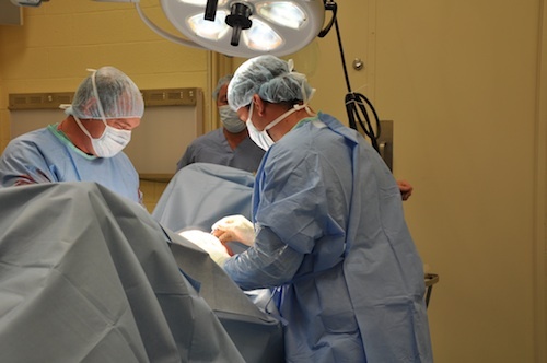
In the event of an emergency, the veterinarians and surgeons of Palm Beach Equine Clinic are available 24/7 to offer the very best care for your equine partner. Palm Beach Equine Clinic is renowned for its referral full-service surgical center and intensive care hospital located in the heart of Wellington, Florida. Board-Certified surgeons, primary care veterinarians, and skilled hospital technicians are available to treat, monitor, and care for critical cases. With world-class veterinarians and a full staff of highly trained technicians, both clients and patients of Palm Beach Equine Clinic are in the best hands possible.
Surgical Capabilities
Palm Beach Equine Clinic offers the latest in technology as the surgical techniques are less invasive and result in faster recovery times for your horse. The surgical team leader, Dr. Robert Brusie, is a nationally renowned, Board-Certified surgeon. Dr. Brusie’s surgical specialties include orthopedic, arthroscopic, and emergency cases. Dr. Brusie has been the head surgeon with PBEC for the past 20 years.
“In the last ten years, colic surgery has come a long, remarkable way,” Dr. Brusie stated. “With our clients, if the horse needs to go to surgery, we get an approximately 95% success rate. We attribute that to the client’s excellent care of their horses, as well as their knowledge to contact us immediately. That being said, colic surgery is always the last resort. We try to help all horses improve medically first.”
Palm Beach Equine’s surgical suite and staff is prepared to handle all types of emergencies, day and night. The large team of 40 veterinarians includes three Board-Certified Surgeons who rotate on-call duties so every day is covered. This aids Palm Beach Equine Clinic veterinarians and all of Southeast Florida with the ability to treat their emergencies requiring surgical assistance as quickly as possible. The state-of-the-art intensive care hospital is equipped with top-of-the-line medical equipment, including digital video cameras for the clinicians to easily monitor their patients from any location, at any time.
It’s no secret that in nearly any medical condition, early diagnosis can lead to a better prognosis – and colitis in horses is no exception. The inflammation of the colon that defines colitis can be fatal, although fortunately, with the proper detection of symptoms and immediate treatment, a positive outcome and recovery far outweigh a negative ending.
Understanding colitis – the symptoms, diagnostics, and treatment— can help in recognizing the condition. Palm Beach Equine Clinic’s Dr. Selina Watt has helped provide some fundamental information that horse owners and barn managers should be aware of in regards to equine colitis.
Understanding Colitis and Its Causes
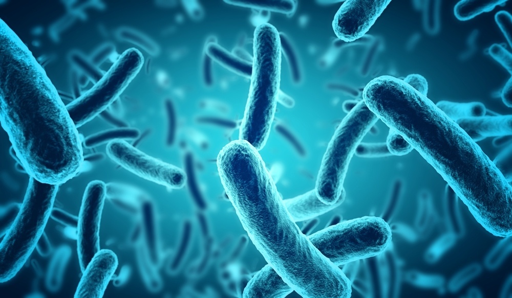
Located in the equine hindgut is the large colon, where microbial digestion and water absorption occurs. The large colon averages 12 feet in length and can hold approximately 20 gallons of feed material and water. When the colon becomes inflamed, the horse is diagnosed with colitis.
While the general definition of colitis is simple and straightforward, the causes can be broader. However, two of the most prevalent causes of colitis are bacterial infections or overuse of medication. Colitis from infectious bacteria is often caused by agents such as Salmonella, Clostridium difficile, or Neorickettsia risticii (Potomac Horse Fever). The non-infectious, right dorsal colitis is often related to the use of non-steroidal anti-inflammatory drugs such as phenylbutazone (Bute).
No matter the cause, each form of colitis leads to a similar inflammation of the large colon. The inflamed colon causes the horse to have diarrhea, as the colon is unable to properly perform its job of adequately absorbing water, electrolytes, and nutrients from the intestinal content. As the condition progresses, leaky membranes of the colon may cause a release of toxins into the bloodstream and the horse will suffer a loss in protein levels. This condition can ultimately affect the entire body as bacteria and toxins circulate, potentially leading to laminitis, founder, protein deficiencies, and a greater risk of complications or lack of a full recovery.
Symptoms and Diagnostics
The first and most conspicuous symptom of colitis is diarrhea. If diarrhea persists, horses can begin to show signs of dehydration and protein loss due to the volume of fluids and nutrients being excreted. Keeping a watchful eye on the consistency of your horse’s manure can be key to catching this condition early. Fever or a lack of energy or appetite may be indicators of colitis and it is recommended to not wait to see what develops but to rather contact a knowledgeable veterinarian for proper diagnostics right away.
Once the horse is under the care of a veterinarian, one of the first things that should be done is bloodwork. In the case of colitis, bloodwork will show decreased white blood cells and protein levels. The severity of the results will indicate how advanced or severe the condition may be. The horse will also generally present with an elevated temperature, and a diagnostic abdominal ultrasound will likely show thickening of the intestinal wall.
Following the initial diagnosis of colitis, a fecal sample is sent to a laboratory where it is tested and analyzed for various forms of bacteria. Comprehensive laboratory results will determine whether the colitis case is infectious or non-infectious. Non-infectious cases can also be diagnosed based on the horse’s history, such as if the horse has been administered Bute for a prolonged period of time.
Treatment and Prognosis
Horses affected by colitis generally require hospital admittance, as they will need fluid therapy and gastro protectants to aid the intestinal wall. If the colitis is caused by infectious bacteria, the patient will also require antibiotic treatment and proper biosecurity measures to prevent transmission. If the bloodwork indicates low protein levels, plasma therapy may also be necessary.
At Palm Beach Equine Clinic, the intensive care management team consists of veterinarians and hospital staff available 24 hours a day, seven days a week. Equine colitis cases cannot simply be administered fluids and left to improve, instead, they require careful monitoring around the clock. If the veterinarian feels the colitis case is severe, the horse may need hourly assessments. This can be of the utmost importance, as colitis cases often rapidly deteriorate without proper veterinary monitoring and swift care.
There is no guaranteed prevention plan for colitis, however, careful management of non-steroidal anti-inflammatory drugs and optimal nutrition can help minimize a horse’s risks of developing the non-infectious colitis condition. With early detection, diagnosis, and proper treatment, equine colitis patients present a positive prognosis.
To ensure the health of your horse, the veterinary team at Palm Beach Equine Clinic is available 24/7. Speak with a Palm Beach Equine Clinic veterinarian regarding the proper medication and nutritional needs of your unique horse by calling 561-793-1599.
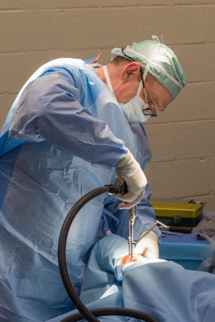
At Palm Beach Equine Clinic in Wellington, FL, the team of Board-Certified surgeons are experts in minimally invasive surgical techniques, aiming to reduce joint disease, resolve lameness, and improve the longevity of sport horse careers.
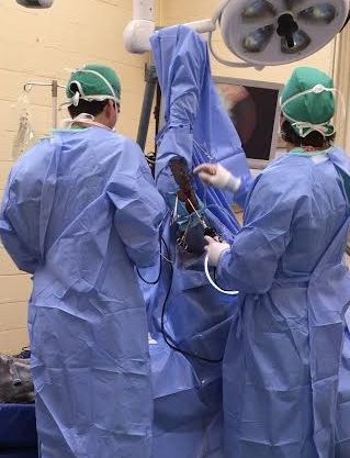
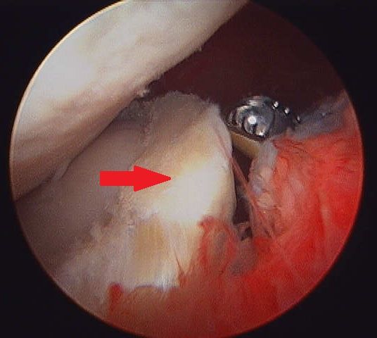
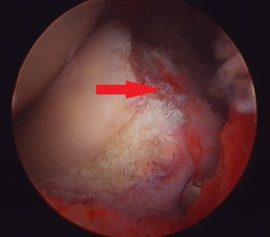
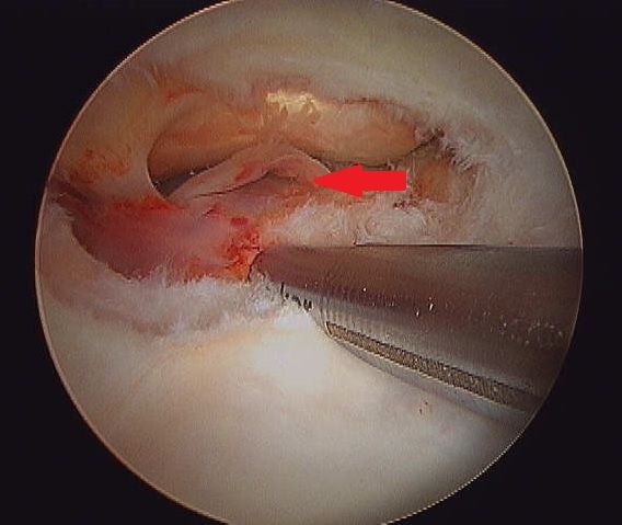
Arthroscopy (or arthroscopic surgery) is a minimally invasive surgical technique that can be performed on an injured joint or synovial structure to accurately explore and treat pathology. The surgery generally involves two very small (8mm) keyhole incisions. The first incision is where the surgeon will insert the arthroscope, which is an instrument with a small surgical grade camera installed that allows a complete, clear view of the interior joint surface. The second small incision is created to insert the surgical instrument to perform the procedure.
Arthroscopy is used to treat a broad range of injuries inside of a joint. Chip fracture removal is a procedure that is particularly commonly in both young Warmblood horses with developmental disease and in racehorses travelling at high speeds. A small chip fracture can cause persistent irritation in the joint as well as arthritis if left untreated. It is best removed immediately so that no further damage is created. The surgeon can go into the joint, remove the chip, and clean up the surrounding cartilage. Most horses recovery quickly and return to their normal athletic activity.
Board-Certified Surgeon Dr. Weston Davis performs many arthroscopic surgeries at Palm Beach Equine Clinic alongside fellow surgeons Dr. Robert Brusie and Dr. Jorge Gomez.
“In many horses, we consider arthroscopy as a prophylactic measure, intervening after injury, but before the development of a generalized degenerative arthritic cycle ensues,” Dr. Davis stated. “Arthroscopy is definitely something that you want to do early in the game if you feel like the horse has joint disease, or a chip, or cartilage disease, or an undefined injury that is not responding appropriately to medical therapy. Arthroscopy can be curative for some of these horses. But if you do not intervene early on in the course of the disease and there is already advanced arthritis, then you have missed your window.
“Arthroscopy is a preferred treatment because it is minimally invasive so most horses can go right back to work,” Dr. Davis continued. “In a typical scenario, we thoroughly explore the joint with the arthroscopic camera, we remove a chip or repair a lesion, and the horse is not lame after the surgery. Because of the small incisions, there is minimal aftercare and horses are often able to go back to work quickly.”
Other common indications for arthroscopic surgery are meniscal disease in the stifle, subchondral cystic lesions, primary cartilage lesions, and debridement of damaged tendinous/ligamentous tissue (such as deep digital flexor tendon tears in the navicular bursa). The surgeons at Palm Beach Equine Clinic can perform arthroscopy on virtually any joint in the horse. Anything from the Temporomandibular Joint (TMJ) of the head to the navicular bursa within the hoof capsule can be explored and treated with this minimally invasive approach.

Almost all arthroscopies are performed under general anesthesia with the horse on its back. New renovations at Palm Beach Equine Clinic include a set of stocks of adjustable height adjacent to a surgeon’s pit, allowing the surgeons to have eye-level access to the joint they are working on, enabling many new procedures on the legs of standing horses.
Minimally invasive surgery allows for a simple and quick recovery for the horse. The traditional horse would be on stall rest with a bandage on until the sutures come out at two weeks, and then start doing some light hand walking and physical therapy. Barring severe damage in the joint or associated tendon/ligament disruption, most cases will undergo a six-week rest and rehabilitation protocol, then return to normal work.
As always, the advanced diagnostic imaging at Palm Beach Equine Clinic permits the surgeons to get a complete evaluation of an injury involving a joint to ensure the best possible outcome. Depending on the injury type, digital radiographs, ultrasound, MRI, and Nuclear Scintigraphy, or a combination thereof, may be used for pre-operative diagnosis and planning. Ultrasound and digital radiography are available for intra-operative use. Intra-operative CT scanning will also be available in the future with the new additions at Palm Beach Equine Clinic.
“When you are inside the joint with an arthroscopic camera, you have the most complete picture of the surface and health of that joint,” Dr. Davis noted.
Diarrhea can be a common problem for horse owners, but how do we know when it is serious? What are some of the causes? How do we treat severe cases and what potential complications you should watch for? Internal Medicine Specialist Dr. Peter Heidmann of Palm Beach Equine Clinic in Wellington, FL, has the answers to these questions and more.
Diarrhea, defined as loose stools, or excessive and overly-frequent defecation, occurs when the intestine does not complete absorption of electrolytes and water. Simple changes in feed, exposure to lush grass, or a bite of moldy hay can cause brief irritation of the bowel, giving a horse diarrhea for a day or two, but anything more than that could be from a variety of more serious causes. Bacteria, viruses, and toxins are all factors that can damage the lining of the bowel and lead to equine diarrhea and other complications.
Causes of Equine Diarrhea
The organisms that cause equine diarrhea are mostly bacteria –Salmonella and Clostridium difficile are among the most common. Clostridium difficile is associated with antibiotic use in both people and horses. While antibiotics are useful to kill bad bacteria, they can also kill good bacteria at the same time, upsetting the balance of flora in the body. If a horse goes on antibiotics for any reason, such as a wound or an infection, that can upset the good bacteria in the intestines and cause bad bacteria, such as Clostridium difficile, to grow.
Clostridium difficile can be found naturally in the environment. There are various types of Salmonella, most adapted to birds or to cattle or other livestock, so horses that are around livestock have a higher rate of becoming infected with that particular bacteria. Horses can also carry Salmonella and not have any symptoms, so they can pass it to each other. If the healthy flora in the horse’s body is thrown off by even a small change in diet, or something bigger like a colic episode, or antibiotics, then Salmonella can grow up in its place.
Another bacterial cause of equine diarrhea can be a disease called Potomac Horse Fever. A bacteria called Neorickettsia risticii, which is carried by snails and conveyed by flies like caddis flies, causes Potomac Horse Fever. For this reason, horses that live near rivers or streams can become infected. During warm weather months, caddis flies pick up the bacteria from the streams and can transfer the disease to nearby horses that accidentally eat the flies or larvae. There are hotbeds for Potomac Horse Fever throughout the U.S., including the Potomac basin where it was first described, as well as many parts of the East Coast, and areas of Oregon, northern California, and Montana.
A viral cause of equine diarrhea commonly seen is Coronavirus. This gastrointestinal virus shreds the intestinal lining and can cause horses to become very sick. The body has to reline the bowel, and it does so quickly, but it takes three to five days, during which the horse may have severe diarrhea and secondary infections.
“Coronavirus was thought for a long time to just be an opportunistic infection and that the virus would take advantage of the horse already being sick, but now it is more and more believed to be the cause of its own type of disease,” Dr. Heidmann stated. “Like all of these diseases, it causes damage to the lining of the bowel and supportive care must be used to help the horse heal. Unlike bacterial infections, however, you cannot directly treat the organism, since there aren’t appropriate drugs to directly treat coronavirus in horses.”
Outside of the infectious causes of equine diarrhea, there are mechanical causes, such as ingestion of sand, which can be a common problem in locations like South Florida. Sand is irritating to the lining of the bowel and can cause damage from its weight, as well as its abrasiveness. In general, sand is irritating enough that the body cannot retain the fluid that it needs in the intestines. As a result, it will cause secretory diarrhea where too much water is being lost. Clearing the sand usually solves the problem and the bowel is then able to reestablish a healthy lining.
A final cause of diarrhea in horses is toxins. Toxic plants, such as Oleander, can be fatal in large doses, but if ingested in small amounts, can be a severe irritant to the bowel. Other toxins that a horse can ingest in the environment, such as phosphate or insecticides, may also cause diarrhea.
Treatment for Equine Diarrhea
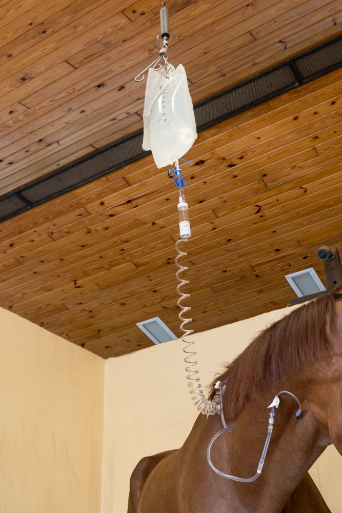
The single most important treatment for diarrhea, no matter the cause, is supportive care. Supportive care includes providing intravenous fluids to replace the fluids lost, providing protein in the form of plasma for the protein lost due to lack of absorption, as well as balancing electrolytes.
The next most important step is taking measures to either reestablish good flora within the gut or to remove the bad bacteria. In the past, a powdered charcoal was used, which is great for absorbing bacteria, but does not absorb the water. A gastrointestinal health supplement called BioSponge® came on the market in the early 2000s through the company Platinum Performance. The product is a purified clay powder that binds the toxins, and also binds the water, so that the horse loses fewer fluids in their equine diarrhea.
While absorbing the bad bacteria and toxins is important, also providing good bacteria in the form of probiotics can be very helpful.
“Probiotics are very variable in their efficacy, but there are some bacteria that are known to be associated with gut health,” Dr. Heidmann noted. “The good bacteria in people, and in horses, that has the most data for being helpful is Saccharomyces Boulardii. Old-fashioned brewers yeast is also Saccharomyces, but it is a different species, Saccharomyces cerevisiae.
“One of the best ways to re-establish healthy flora is Transfaunation, which is taking a healthy horses manure, filtering it, and then tubing it into the sick horse,” Dr. Heidmann added. “That is one of the most dramatic treatments out there. It provides the good ‘bugs’ that the horse is losing through the diarrhea. You will often see foals eating their mother’s manure. It is an instinctual habit to get the good bugs into their stomach. We only do that in the sickest of cases. Whatever the route, it makes a big difference to provide the good bugs because that creates the environment for the gut to heal.”
While some antibiotics are warranted in the right situation, Dr. Heidmann pointed out that they are not necessary as often as people would think.
“With people or dogs, if we get Salmonella or some other intestinal infection, we almost always go on antibiotics, but because antibiotics are the cause of many cases of colitis in horses, in general, that is not the best strategy,” Dr. Heidmann stated. “There are a couple of exceptions. Clostridium difficile does respond to antibiotics, metronidazole being the most common one. For Potomac Horse Fever, Tetracycline broad-spectrum antibiotics are the best.”
Biosecurity measures should also be taken to protect healthy horses from an infectious barn-mate. Dr. Heidmann recommends complete isolation of the sick horse while it is ill, and for a minimum of two full weeks after the infection has been clinically resolved. This includes no horse-to-horse contact, as well as no shared use of wheelbarrows, pitchforks, etc.
Molecular and DNA testing can be done to make sure that the horse is infection-free, however, Dr. Heidmann warns that testing can be problematic.
“There is a very high number of false negatives, meaning there is truly some infection there, but the lab cannot find it,” Dr. Heidmann stated. “There can be times when the horse is shedding bugs, but the tests do not pick it up. The state-of-the-art standard of care is a DNA test called ‘PCR’, and yet you still have to do multiple tests to get a positive test and get a diagnosis. Still, the best way to be safe is to continue testing until you are sure.”
Complications of Equine Diarrhea
Dr. Heidmann warned of common complications in severe diarrhea cases, laminitis being highest on the list. With the sickest of horses, it is unfortunately not uncommon for the veterinarian to get the gut fixed over three to five days, and then find that the feet have started to become very inflamed due to toxins in the bloodstream. If the horse loses the lining of its intestine, then the good and bad bacteria that are supposed to be contained in the intestine can “leak” out into the bloodstream and are free in the abdomen. Those bacteria are then dying either from an attack by the immune system or antibiotics, and they release endotoxins into the bloodstream, which along with other inflammatory products, can cause laminitis.
Another serious complication is blood clotting. The sick horse may become very low on blood protein when the bowel lining is damaged, which can cause clotting abnormalities. The horse may have difficulty clotting or they may become prone to abnormal increases in clotting. The horse might seem better, and then it will develop a clot somewhere in the body. It can be anywhere, but it is most often in the intestine itself, which is usually fatal. In general, horses like this are treated with supplemental protein in the form of plasma. In some cases, the veterinarian will also provide anticoagulant medications.
Although some cases of equine diarrhea are brief and easily resolved, Dr. Heidmann reminds that serious cases can go downhill fast, and it is important to refer to an expert.
“The biggest sign of a problem is duration,” Dr. Heidmann concluded. “If it is one day, it could be that they had a bite of bad food or something simple. If there are fevers or lethargy, those are instant warning signs. If it lasts for days, or if they go off their feed, those are instant warning signs. That is when you should call your veterinarian right away, especially because as they start to go downhill, these complications really amplify. The worst cases are the ones that have been smoldering for a day or two.”
Dr. Heidmann and the veterinarians at Palm Beach Equine Clinic are always available and encourage owners to contact the clinic at the first sign of a problem.
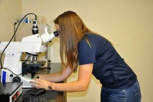
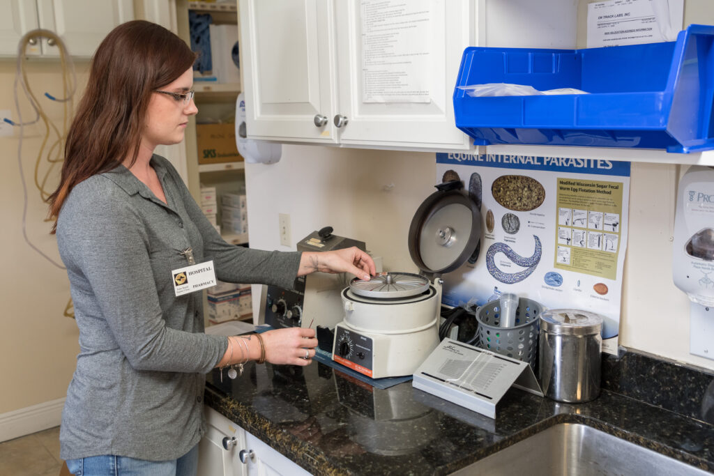
Palm Beach Equine Clinic, based in Wellington, FL, boasts one of the most advanced laboratory facilities in the country with onsite equipment capable of performing internal hematology, chemistry, and microbiology testing, as well as many regenerative therapies.
The facilities provide a plethora of services that are not only useful to Palm Beach Equine Clinic veterinarians, but also the many veterinarians who visit South Florida during the winter show jumping, dressage, and polo seasons.
Functions of the Palm Beach Equine Clinic Laboratory
Coupled with technologically advanced imaging services, state-of-the-art surgical capabilities, and care from renowned veterinarians, Palm Beach Equine Clinic takes pride in their onsite laboratory, which offers vital services to a range of clients providing rapid results.
Most commonly used, hematology is the study of blood, its chemistry, and components. A complete blood count or CBC determines the number and type of white and red blood cells circulating through the bloodstream. This can be quickly and easily performed in Palm Beach Equine Clinic onsite laboratory. Changes in these blood cells can indicate inflammation, infection, or disease. Quick diagnosis leads to more proactive and efficient treatment plans.
A clinical chemistry is the study of the chemical composition of a sample. Typically, the liquid portion of blood (either serum or plasma) is used for testing components such as electrolytes, kidney enzymes, and muscle enzymes. The serum or plasma is evaluated to determine the efficiency and health of specific organs.
Finally, microbiology is the study of small organisms such as bacteria, viruses, fungi, and other single-celled life forms. Hundreds of microbiology tests can be performed at Palm Beach Equine Clinic to look for signs of infection. The cultures are used to identify a specific bacteria or fungus present and sensitivity tests are used to determine which treatment, such as an antibiotic, will most effectively treat the infection.
For Palm Beach Equine Clinic veterinarian Dr. Samantha Miles, having an on-site laboratory with many different services enables her to provide faster and more affordable results to her clients and the horses in her care.
“We tend to get results so much faster in our own lab,” said Dr. Miles. “Also, an in-house culture is much less expensive than sending the sample away. It takes 24 hours to run a culture and 24 hours after that for the sensitivity. So usually it takes 48 hours to get a full culture and sensitivity, whereas to send that away you’re looking at least at 72 hours minimum and sometimes it’s a couple days longer than that.”
While laboratory technologies are common in determining a diagnosis and identifying different infections and viruses; they can also be used to more effectively and quickly treat common equine problems such as colic.
“There has been a lot of research lately comparing blood lactate to the abdominal fluid lactate, and the difference being a good indicator of whether a colic is surgical versus medically managed,” said Dr. Miles. “Sometimes it’s obvious, but not always so it is really helpful to have that capability. What we do is test a blood lactate sample using a lactometer, which takes about a minute. If a horse is dehydrated and has a higher lactate in the abdomen, we will rehydrate the horse and take it again. If the lactate value doesn’t decrease after rehydration we have a good indication that it is a real number and there is a surgical problem.”
Regenerative Therapies
According to Dr. Miles, some of the most impressive functions of the Palm Beach Equine Clinic laboratory include its regenerative therapy capabilities such as:
- Stem Cells
- Platelet-Rich Plasma (PRP)
- Interleukin-1 Receptor Antagonist Protein (IRAP)
- Pro-Stride Autologous Protein Solution (APS)
These can be applied to previously difficult to manage joint diseases and injuries using natural-occurring proteins, cells, and other natural processes originated from within the body of the horse. Essentially, these treatments use the horse’s own biological mechanisms to stimulate healing without the use of steroids or other drugs.
Stem cells are commonly derived from bone marrow which are cultured and multiplied into millions of stem cells. The cultured stem cells are injected into the affected tendon, ligament, or joint to encourage healing. PRP is another byproduct that is internally sourced from blood platelets in a matter of minutes. The platelets are combined with numerous growth factors that are released upon activation and can enhance healthy inflammatory cells in areas of tissue injury, form new blood vessels, new connective tissue, and aid in the regeneration of skin when injected.
IRAP, stands for Interleukin-1 Receptor Antagonist Protein, is used to treat equine athletes that are susceptible to musculoskeletal injuries and osteoarthritis or degenerative joint disease. Joint trauma results in the release of inflammatory mediators such as Interleukin-1 (IL-1). IRAP uses a horse’s own anti-inflammatory protein found within the blood to counteract the destructive effects of IL-1 to slow the process of osteoarthritis. The process works by binding to the IL-1 receptors in the joint and blocking the continuation of damage and inflammation.
“We often see joint damage in sport horses because of the nature of their work, but we try to avoid overuse of steroids in joints because steroids can have long term effects on cartilage,” said Dr. Miles. “This is a way we can manage joint disease and stop inflammation without having to consistently use steroids every time. Some of our clients will maintain their horses on IRAP alone for joint injections.”
Most recently, Palm Beach Equine Clinic has added a Pro-Stride Autologous Protein Solution (APS) Device, which is a new up and coming treatment that combines PRP and IRAP treatments but provides faster results.
Pro-Stride APS will reduce pain associated with arthritis and deliver anti-inflammatory proteins capable of slowing cartilage damage and improving mobility through the Interleukin-1 Receptor Antagonist Protein. The process can provide pain relief for up to one year following a single injection, which includes 20 minutes of blood processing in the Palm Beach Equine Clinic laboratory with no incubation period.
“I believe we are learning more about these technologies with more advanced science behind what they do and how they do it, “ said Dr. Miles. “These treatments are natural, drug-free, competition safe and necessity drives the need for regenerative therapies in the sport horse world.
“It’s all these new regenerative therapies that I think make our lab more state-of-the-art,” continued Dr. Miles. “They set us apart and are also tools that referring vets can make use of. The bottom line is that we have the ability to get horses back significantly faster after injury by using these therapies.”
An Expert Team
The laboratory at Palm Beach Equine Clinic offers 24-hour service with quick and efficient processing of blood work and test results. While veterinarians, or interns under the supervision of a veterinarian, are involved in a lot of the laboratory processing, the Palm Beach Equine Clinic laboratory is also staffed by 24-hour technicians. As a result, test results are returned to veterinarians and subsequently horse owners as fast as possible.
“We are lucky enough to have access to the technologies found in the Palm Beach Equine Clinic laboratory and work with people who have the experience, knowledge, and training to run such an advanced facility,” said Dr. Miles. “We always look forward to welcoming new and returning referring vets to the equipment, technology, and innovation that we have available at Palm Beach Equine Clinic. We take pride in our symbiotic relationship with veterinarians visiting Florida from around the country and the world.”
Palm Beach Equine Clinic of Wellington, FL, proudly offers advanced services to referring veterinarians and clients in equine podiatry with the expertise of Dr. Stephen O’Grady. As the show season continues on, some horses may be experiencing foot soreness or new lameness that could be related to their farriery.
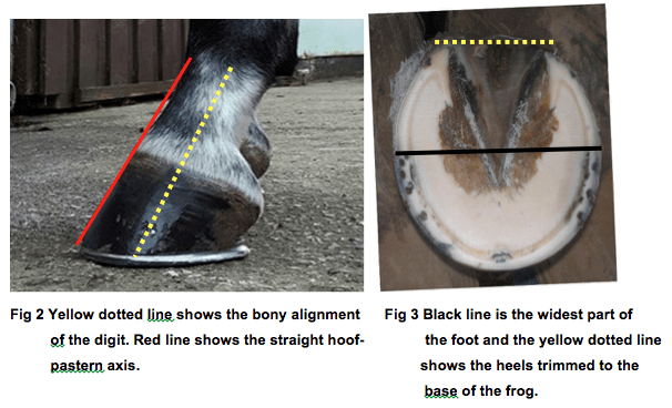
The importance of high quality hoof care in the competition horse can’t be denied. The equine hoof is unique, as it is comprised of a group of biological structures that follow the laws of biomechanics (Figure 1). The farrier is a major asset during the show season as he or she can be proactive in maintaining the health of your horse’s feet and thus preventing lameness.
There are three very important aspects of farriery science that the farrier will use to keep your horse sound, which are trimming the foot in conjunction with the size and placement of the horseshoe. Typically, a farriery session will begin with an evaluation of the conformation of each hoof from the front, side, and behind to observe the height of the heels. Next, the farrier should observe the horse in motion to see whether the horse’s foot lands heel first, flat or toe first. All this information is considered and evaluated before the farrier begins shoeing.
Trim
Regarding the trim, many farriers no longer use the term ‘balance the foot’ – which has no meaning – and have begun to use guidelines or landmarks when approaching the trim. The guidelines used are trimming to achieve a straight hoof-pastern axis, using the widest part of the foot which correlates to the center of rotation, and trimming the palmar foot (heels) to the base of the frog or to the same plane as the frog (Figure 2, 3).
A closer look at these three guidelines, which are all interrelated, will help to show their importance. If the dorsal (front) surface of the pastern and the dorsal surface of the hoof are parallel or form a straight line, then the bones of the digit (P1, P2, P3) are in a straight line, and the force from the weight of the horse will go through the middle of the joint. Furthermore, and equally important, if the hoof-pastern axis is straight, the weight will be distributed evenly on the bottom of the foot.
The second guideline is the center of rotation (COR), and as the COR is located a few millimeters behind the widest part of each foot, it allows the farrier to apply appropriate biomechanics to each foot. The foot is trimmed in approximate proportions on either side of the widest part of the foot, which provides biomechanical efficiency.
Heels
Lastly, one should trim the palmar section of the foot to the base of the frog or trim such that the heels of the hoof capsule and the frog are on the same plane. Adherence to this guideline keeps the soft tissue structures (frog, digital cushion, ungula cartilages) within the hoof capsule, which are necessary to absorb concussion and dissipate the energy of impact.
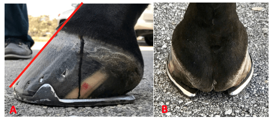
We must remember that heels do not grow tall, they grow forward. If we allow the heels to migrate forward, the soft tissue structures will be forced backward out of the hoof capsule. Furthermore, as the heels migrate forward, the weight is placed on the bone and lamellae, thus bypassing the soft tissue structures of the foot. Allowing the heels to migrate forward also decreases the ground surface of the foot. An example of this guideline is shown in Figures 4A & 4B, where the palmar foot was trimmed appropriately and a size larger shoe was applied to properly distribute the weight.
These three guidelines can be applied to any foot and they serve as a basis for maintaining a healthy foot and a basic starting point for applying farriery to a horse with poor foot conformation or one with a distorted hoof capsule. Figures 5A & 5B will illustrate a hoof where all three of these guidelines have been applied.
Farriery and Horse Showing
Many horses are given a rest from competition, which includes their feet, following a heavy competition load such as Wellington’s Winter Equestrian Festival and Adequan Global Dressage Festival. Many horses arrive with very reasonable foot conformation, but upon arrival the farriery can change and many horses are shod with various specialty shoes, wedges, pads, pour-ins, etc. as a means of protection, and perhaps, to enhance their performance.
As the season progresses and the workload increases, the sole thickness starts to decrease and the feet become softer from multiple baths; now the farriery that was applied for protection may be causing pressure on the thinner, softer structures of the foot, thus becoming somewhat detrimental. Furthermore, the horses continue to be trimmed and shod on a monthly basis and the change in the integrity of the hoof structures without investigating can cause horses to be over-trimmed. Additionally, as the season starts into March, the structures of the foot deteriorate further as a result of the workload, and many horses become foot sore. At this point, the farrier options are limited because they may have been used at the beginning of the season.
Luckily, Palm Beach Equine Clinic offers a farriery consultation service by Dr. Stephen O’Grady to both veterinarians and farriers. This unique service provides a second opinion or ‘another set of eyes’ to both professions when treating difficult farriery cases for ideas on other options to help these foot sore horses.
Palm Beach Equine Clinic provides experience, knowledge, availability, and the very best care for its clients. To find out more, please visit www.equineclinic.com or call 561-793-1599.
Palm Beach Equine Clinic (PBEC), located in Wellington, FL, offers advanced diagnostic imaging, world-renowned surgical talent, and state-of-the-art facilities necessary to quickly diagnose, treat, and repair horses with condylar fractures, making PBEC one of the leading facilities in the U.S. for condylar fracture repairs.
With thanks to the technology required for early diagnosis and experienced surgeons on staff, horses that are admitted to PBEC for condylar fracture repairs are more likely to return to training quickly. Most commonly seen in Thoroughbred racehorses and occasionally polo ponies or eventing horses, a condylar fracture was once considered a career-ending injury. Today, however, advances in technology aid in a full recovery with horses regularly returning to competition in their respective divisions.
What is a Condylar Fracture?
A condylar fracture is a repetitive strain injury that results in a fracture to the cannon bone above the fetlock due to large loads transmitted during high-speed exercise. On a radiograph, a condylar fracture appears as a crack that goes from the fetlock joint up the cannon bone. Lateral fractures many times exit the bone usually one-third of the way up the bone. Medial fractures will oftentimes spiral up to the hock or knee. Medial fractures are much more common in the hindlimb than the forelimb. It is the spiral fractures that are more difficult, due to the fact that the extent of the spiral cannot be identified radiographically. If the surgeon cannot identify the fracture, then that part of the fracture cannot be repaired.
“A condylar fracture is a disease of speed,” said Dr. Robert Brusie, a surgeon at PBEC who estimates that he repairs between 30 and 50 condylar fractures per year. “A fracture to the left lateral forelimb is most common in racehorses as they turn the track on a weakened bone and increased loading on the lateral condyle.”
Condylar fractures are further categorized into two classes. An incomplete and non-displaced fracture means that the bone fragment is not separated from the cannon and is still intact with its original position. A complete and displaced fracture means the fragment has detached from the cannon bone and this fracture can often be visible under the skin. Displaced condylar fractures have a somewhat lower prognosis due to the fact that soft tissue structures, such as the joint capsule, become torn. When these structures heal, they are thicker, which makes the joint less flexible.

“Most lateral condylar fractures are fairly simple for us to fix,” said PBEC surgeon Dr. Weston Davis. “Medial condylar fractures tend to be more complicated configurations because they often spiral up the leg. Those require more advanced imaging and more advanced techniques to fix.”
What is the Treatment?
The first step to effectively treating a condylar fracture through surgery is to accurately and quickly identify the problem. PBEC’s Board-Certified Radiologist Dr. Sarah Puchalski utilizes the advanced imaging services at PBEC to assist in the diagnosis.
“Stress remodeling can be detected early and easily on Nuclear Scintigraphy before the horse goes lame or even develops a fracture,” said Dr. Puchalski. “Early diagnosis of stress remodeling allows the horse to be removed from active race training and then return to full function earlier. Early diagnosis of an actual fracture allows for repair while the fracture is small and hopefully non-displaced.”
Once identified as a condylar fracture, PBEC surgeons step in to repair the fracture and start the horse on the road to recovery. Depending on surgeon preference, condylar fracture repairs can be performed with the horse under general anesthesia, or while standing under local anesthesia and sedation. During either process, surgical lag screws are used to reconnect the fractured condyle with the cannon bone.
“For a very simple and small non-displaced fracture, we would just put in one to two screws across the fracture,” explains Dr. Davis. “The technical term is to do it in ‘lag fashion’, such that we tighten the screws down heavily and compress the fracture line. Many times the fracture line is no longer visible in x-rays after it is surgically compressed. When you achieve good compression, the fractures heal very quickly and nicely.”
More complicated fractures, or fractures that are fully displaced, may require more screws to align parts of the bone. For the most severe cases of condylar fractures, a locking compression plate with screws is used to stabilize and repair the bone.
Severe condylar fractures often require general anesthesia, but for PBEC surgeon Dr. Jorge Gomez, approaching a simpler non-displaced condylar fracture while the horse is standing helps to aid in a faster recovery and successful surgical outcome.

“I think it takes the risk of anesthesia away and is a faster surgery from the time the horse comes in to the time the horse recovers,” said Dr. Gomez. “I will just sedate the horse and block above the site of the fracture. Amazingly, horses tolerate it really well, and it is very convenient for medial condylar fractures. In these cases, the fracture can spiral all the way up through the cannon bone, and they have a tendency to develop complete catastrophic fractures that can happen at any time after the injury. That risk can be significantly increased by the recovery from general anesthesia. Our goal is always to have the best result for the horse, trainers, and us, as veterinarians.”
According to Dr. Gomez, the recovery time required after a standing condylar fracture repair is only 90 days.
While Dr. Brusie, Dr. Davis, and Dr. Gomez are all seasoned in quickly and effectively repairing condylar fractures, PBEC is helping them to stay on the cutting edge of surgical techniques. PBEC is currently renovating its facility with plans to give surgeons a new approach to fix condylar fracture repairs. A set of stocks and surgeon’s pit have been added with the ability to give the surgeon eye-level access to the fracture with the patient standing and subsequently simplifying the procedure by reducing the risk from recumbent recovery.
What is the Prognosis?
One of the most common questions regarding an equine injury is, “Will the horse return to work?” Thanks to advanced imaging and surgical techniques, the answer to that question when involving a condylar fracture is most likely, “Yes.” At PBEC, a condylar fracture diagnosis rarely results in the end of a racehorse’s career.
Diagnostic imaging plays a major role in assisting to diagnose, surgically map, and follow up on condylar fractures. After primary use to diagnose a condylar fracture, digital radiographs are also used after surgery to ensure that a fracture repair was completely successful. According to Dr. Davis, scanning two planes during and after surgery gives a full view of the fracture and repair techniques, immediately indicating the success of the procedure before moving the horse on to recovery.
“A condylar fracture was once considered the death of racehorses, and as time and science progressed, it was considered career-ending,” said Dr. Brusie. “Currently, veterinary medical sciences are so advanced that we have had great success with condylar fracture patients returning to full work. Luckily, with today’s advanced rehabilitation services, time, and help from mother nature, many horses will come back from an injury like this.”
Palm Beach Equine Clinic provides experience, knowledge, availability, and the very best care for its clients. Make Palm Beach Equine Clinic a part of your team!